>Corresponding Author : Michel Leclerc
>Article Type : Research Article
>Volume : 2 | Issue : 2
>Received Date : 06 May, 2022
>Accepted Date : 17 May, 2022
>Published Date : 20 May, 2022
>DOI : https://doi.org/10.54289/JCVR2200106
>Citation : Leclerc M. (2022) Cytotoxicity Comparisons between "Young Protein" from Sea Star Igkappa Gene and Doxorubicine against Cancerous Mcf7 Cells. J Clin Vet Res 2(2): doi https://doi.org/10.54289/JCVR2200106
>Copyright : © 2022 Leclerc M. This is an open-access article distributed under the terms of the Creative Commons Attribution License, which permits unrestricted use, distribution, and reproduction in any medium, provided the original author and source are credited.
Research Article | Open Access | Full Text
Immunology of Invertebrates Orléans University, 556 rue Isabelle Romée, 45640 Sandillon (France)
*Corresponding author: Michel Leclerc, Immunology of Invertebrates Orléans University, 556 rue Isabelle Romée, 45640 Sandillon (France)
Abstract
Some years ago, we found that Axial organ (AO) cells exerted an induced and spontaneous cytotoxicity against SP2 and MBL2 mouse tumoral cells. Recently, we discovered a sea star Igkappa gene with immune properties. This gene was first, inserted in a CMV (cytomegalovirus) and finally in a plasmid called « young » plasmid.
In the present experiment the sea star IGKappa gene was cloned in HeK human cells to produce the specific protein.
The induced« young » protein exerted a weak cytotoxicity against MCF7 cells.
Keywords: Invertebrates; Vertebrates; Cloning; CMV; Hek Cells; MCF7 Cells
Abbreviations: AO: Axial organ, MCF7: Mammal Cancerous Cells
Introduction
In 1983, Luquet and Leclerc [1] shown that the axial organ cells (AO cells), exerted a spontaneous and induced cytotoxicity against mouse SP2 myeloma cells and MBL2 cells.
The AO cells included essentially lymphocytes and phagocytes [1]
40 years later, we discovered a sea star Igkappa gene [2], with immune properties [3]
We have studied the behaviour of the « young » protein secreted by the sea star Igkappa gene, in front of human malignant: A-375 melanome cells, human Osteosarcome cells (U2oS cells) and human malignant cells Hela, by the use of plasmids. In the present time we study the "young" protein issued from HeK cells against MCF7 cancerous cells.
Materials and Methods
a. Mouse SP2 and MBL2 cells were cultured in our laboratory
b. Gene cloning in HeK human cells was at last realized [4]; The protein is actually studied against human mammal cancerous cells (MCF7). A control of cell viability was done with doxorubicine [5].
We incubated the same number of MCF7 cells at passage 31 in a 96 well plate (some wells did not contain cells = background wells) and let the cells grow for 48h, until the confluence was around 70%. Then, the cells were treated with Doxorubicin Hydrochloride and young protein dissolved in DPBS at the doses indicated in the table below (final volume 100uL cell culture media). Some wells were treated with the same volume of DPBS (vehicle controls). All conditions were tested in triplicates. After 24h treatment, the cells were gently washed with DPBS, and 100uL cell culture media was added. For the cytotoxicity analysis, 20uL of the reagent (Abnova kit, reference KA4151) were added to each well, and the reading was performed after 1h, 2h and 3h. The absorbance is measured at 570nm and 605nm. The cell viability is proportional to an increased 570/605nm. The reading was best performed at the 3h timepoint (keep in mind the treatment was 24h long, this is just the reagent incubation time). For the results analysis: the ratio 570/605nm absorbance was calculated for each well, and the background signal (from the wells without cells) was substracted. The PBS-treated cells (Control) were stablished as the 100% viable, and the viability on the rest of the wells was calculated as: %viability = ODsample/ODcontrol*100
Results
Results are as follows as seen in Table 1:
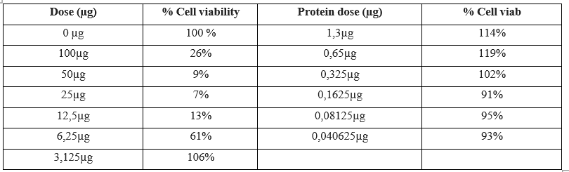
Table 1: Left column: Dose of Doxorubicin in µg and cell viability on MCF7 cells which falls down at 7% for a concentration of 25µg of Doxo
Right column Young protein dose and cell viability which falls down at 91% for 0,1625 µg with "young" protein.
Discussion, Conclusion
The left column of the Table 1 shows clearly that Doxorubicin at a concentration of 25µg exerts a stronger cytotoxic activity than at 0µg or 3,125 µg. On the other hand our young protein issued from Cloning in HeK cells possesses a weaker activity on MCF7 cells, the corresponding cell viability is 91% for a 0,1625µg concentration of young protein. We can expect first, that cloning in
HeK cells has changed the parameters of the young protein against MCF7 cancerous cells but secondly it seems fundamental to test our protein against another type of tumoral cells such as Hela cells
References
- Luquet G and Leclerc M. (1983) Immunol Lett. 6: 107-108. [Ref.]
- Vincent N, et al. (2014) A new gene in A. rubens: A sea star Ig kappa gene. Metagene. 2: 320-322. [Ref.]
- Leclerc M and Otten P. (2014) Immune Properties Corroborated by A. Rubens Sea Star Igkappa Gene. SAJ Biotechnology. 1: 104. [Ref.]
- Leclerc M. (2021) Recombinant Sip Young 6 His Protein Production from the Sea Star Igkappa Gene, after Cloning. Eur J Biol and Biotechnol. 2(2): 85-86. [Ref.]
- Lovitt CJ, et al. (2018) BMC Cancer. 18: 41. [Ref.]