>Corresponding Author : Mark E Peacock
>Article Type : Case Report
>Volume : 6 | Issue : 2
>Received Date : 13 July, 2026
>Accepted Date : 24 July, 2026
>Published Date : 28 July, 2026
>DOI : https://doi.org/10.54289/JDOE2600106
>Citation : Croft W, Ghaly M, Abdelsayed R, Timothius CJC, and Peacock ME. (2026) Surgical Ciliated Cyst of the Maxilla: Case Report and Review of the Literature. J Dent Oral Epidemiol 6(2): doi https://doi.org/10.54289/JDOE2600106
>Copyright : © 2026 Croft W, et al. This is an open-access article distributed under the terms of the Creative Commons Attribution License, which permits unrestricted use, distribution, and reproduction in any medium, provided the original author and source are credited.
Case Report | Open Access | Full Text
1Department of Periodontics, Dental College of Georgia at Augusta University, Augusta, Georgia
2Department of Diagnostic Sciences, Dental College of Georgia at Augusta University, Augusta, Georgia
*Corresponding author: Mark E Peacock, Department of Periodontics, Dental College of Georgia at Augusta University, Augusta, Georgia
Abstract
Surgical ciliated cysts (SCC) are rare benign jaw cysts that are associated with inadvertent transplantation of respiratory epithelial tissue from a previous surgery and/or trauma to the maxillary sinus. This report describes a SCC of the posterior maxilla that occurred 16 years after previous left maxillary sinus surgery for bone augmentation. A 73-year-old male was referred for evaluation of an asymptomatic well-defined unilocular radiolucency in the left posterior maxilla, mesial to #15. The patient’s history is significant for implant insertion in the left side of the maxilla. In 2010, the patient underwent maxillary sinus-lift surgery with implant insertion The patient later developed peri-implantitis and the #14 site fixture was removed, combined with a guided bone regeneration procedure which did not involve the maxillary sinus. Surgical therapy to explore the cyst-like lesion before fixture placement was implemented after consultation, during which the cystic lesion was identified and enucleated. Histopathology, along with the sinus surgery history, was consistent with a diagnosis of SCC of the maxilla. Follow-up has shown no complications and no evidence of recurrence. The patient is scheduled for placement of an implant at #14 site. Though many SCCs present without clinical signs/symptoms and are discovered incidentally, some rarely may appear as large symptomatic swellings with multilocular radiographic findings. Clinicians need to be attentive to the possibility of SCC in their differential diagnosis, particularly when the patient’s history reveals previous jaw surgery involving areas proximate to the maxillary sinus.
Keywords: Surgical Ciliated Cyst; Maxillary Sinus; Jaw Cyst
Abbreviations: SCC: Surgical Ciliated Cysts, PMC: Postoperative Maxillary Cyst, SCCM: Surgical Ciliated Cyst of the Maxilla, GBR: Guided Bone Regeneration
Summary
This case report documents a rare cyst of the maxilla, the surgical ciliated cyst, that occurred in an older individual in a site where 16 years earlier he had a maxillary sinus lift surgery performed. Practitioners should be attentive to suspected cystic pathology, especially in patients who have previously had surgery/trauma involving or near the maxillary sinuses.
Introduction
Surgical ciliated cysts are uncommon benign cystic lesions in jaw bones whose etiology has been shown to be from previous trauma/surgery near the maxillary sinus. The accidental entrapment of respiratory ciliated epithelium into the maxilla or mandible from previous sinus surgery is the chief distinguishing feature. A postoperative maxillary cyst (PMC) was first described in the literature in 1927 by Kubo in Japan, [1] which later in 1958 was coined as the surgical ciliated cyst of the maxilla (SCCM) by Gregory and Shafer [2]. SCC was recently (2022) added to the World Health Organization (WHO) Classification of Head and Neck Tumours [3]. Incidence of SCC in western countries is not very common, reported to be roughly 1.5% of oral cysts [4]. However, the occurrence rate in Japan has been shown to be much higher, 16 to 19.5% of gnathic cysts [5,6]. It has been hypothetically proposed that the higher incidence in Japan was attributed to the higher rate of surgical treatment of chronic sinusitis in young Japanese adults, but, current thought is that genetic anatomical factors may be involved.
The usual age group that SCCs are observed are 50s to 60s, and the period of time between diagnosis and the earlier assumed causal surgical procedure can be 20 years [7]. Conversely, Kahn et al in 2021 published findings that the interval for cyst development after maxillary sinus floor augmentation before dental implant placement was only 0.5 to 10 years [8]. The most common location where SCCs occur is the maxilla, but can also be rarely found in the mandible [9]. Recently Brisset et al in a systematic review of mandibular SCC case reports revealed that they are located mainly in the anterior mandible, due chiefly to the type of surgical procedure itself [10]. Their reported patients were usually treated previously by genioplasty together with nasal autograft, or genioplasty and maxillary osteotomy. In order to prevent the accidental implantation of sinus mucosa, the authors proposed using a different procedure, such as sliding genioplasty, which does not employ nasal grafts [10]. SCCs can present as asymptomatic without swelling or painful enlargements that may affect teeth in the site. Radiographically the lesions are usually unilocular, but some larger cysts can be multilocular [11]. Most studies do not report any major difference in incidence in male vs female patients. The recommended treatment for most SCCs is curettage and enucleation, and the prognosis is usually good. Some studies report a recurrence rate between 6 and 20% [12,13]. A possible reason for return of the cyst could be incomplete removal of the epithelial tissue lining, which could also migrate to nearby bone [14].
Even though SCCs are still considered relatively rare, surgeons should still be suspicious and include this in the differential diagnosis in any patient who had previous maxillofacial trauma and/or surgical procedures near maxillary sinuses. This case describes a patient who 16 years earlier had undergone a direct sinus window surgical procedure to lift the Schneiderian membrane and augment osseous tissue for dental implant placement. Subsequently a possible cyst-like radiolucency was discovered upon specific consultation/examination of the patient for placement of implants in the upper left posterior maxilla. Long-term follow-up should be mandatory for any patient who has had previous surgery in the maxillofacial region that possibly involved the antral region(s).
Case Report
The patient, a 73-year-old African-American male, was referred and treated at the Department of Periodontics, the Dental College of Georgia (DCG) at Augusta University (AU), Augusta, Georgia for consultation/exam/treatment for possible dental implant placement in the left posterior maxilla. A review of the medical history was significant for transient ischemic attack (1986), hypertension, atrial fibrillation, and benign prostatic hyperplasia. Current medications included lisinopril, amlodipine, metoprolol, tamsulosin, rivaroxaban, flecainide, and multivitamins. There were no known drug allergies. A comprehensive exam revealed that the patient had previously been treated for insufficient bone height in the left posterior maxilla with a direct sinus lift plus dental implant placement at #14 site (2010). Due to severe peri-implantitis, the implant was removed and a guided bone regeneration (GBR) was completed in early 2025 by a previous practitioner. Radiographic (cone beam computed tomography, CBCT) exam disclosed a well-demarcated unilocular radiolucency measuring approximately5 x 5 mm mesial to tooth #15 (Figures 1 and 2). The decision was made to explore and biopsy the possible lesion before any permanent fixture was placed in the area. Parenteral sedation was achieved with midazolam, fentanyl, and diphenhydramine. Local anesthesia was obtained with topical 20% benzocaine plus 2% lidocaine with 1:100,000 epinephrine, 4% articaine with 1:100,000 epinephrine, and 0.5% bupivacaine with 1:200,000 epinephrine. A buccal mucoperiosteal flap was elevated in the posterior maxilla, and the cyst was identified. No osseous perforation was present. A bony window was prepared, and the cyst was curetted and removed (Figure 3). Thin cystic soft fragments (Figure 4) were removed and submitted for pathological examination. Primary closure was obtained at the surgical site (Figure 5).
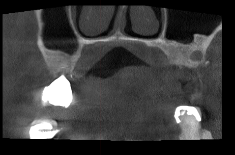
Figure 1: CBCT showing unilocular lesion posterior left maxilla
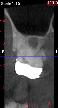
Figure 2: Closeup view of CBCT
Pathological Findings
Microscopic examination revealed sections composed of a thin fibrous capsule lined with respiratory epithelium consisting of ciliated pseudostratified columnar epithelium were noted (Figure 6, A and B). A diagnosis of surgical ciliated cyst of the maxilla was made. Healing was within normal limits and uneventful. The post-surgical follow-up (four months) so far has revealed no recurrence.

Figure 3: Surgical view of cyst curetted and enucleated

Figure 4: Remnants of removed lesion before histology submission

Figure 5: Surgical site sutured with primary closure obtained
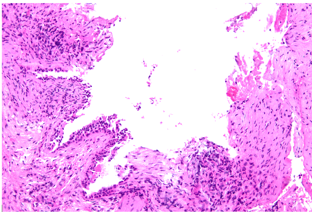
Figure 6A: Medium-power photomicrograph (hematoxylin and eosin [H&E], x 20) showing a fibrous capsule →lined with ciliated columnar epithelium
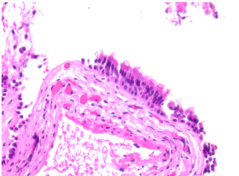
Figure 6B: High-power photomicrograph (hematoxylin and eosin [H&E], x 40) showing a fibrous capsule lined with ciliated columnar epithelium
Discussion
The surgical ciliated cyst is an uncommon lesion in American-European countries, whereas in Japan the incidence approximates up to 20% [15]. It has been suggested by Hii et al that the SCC may be underreported in populations outside of Japan, especially now since in 2022 WHO added the lesion to the Classification of Head and Neck Tumours [16]. While some SCCs may present with signs and/or symptoms, many are asymptomatic and are unexpectedly found upon routine examination [17]. Mandatory for a diagnosis of SCC to be made is the combination of histology showing respiratory epithelium plus a surgical/trauma history involving the maxillofacial region. A portion of the sinus epithelium from a previous procedure is possibly reintroduced into adjacent bone, and over time the tissue grows and forms a cyst [18,19]. Recurrence is very rare, but long-term follow-up should still be required.
Other pathology that could be considered in the differential diagnosis are radicular cyst, odontogenic keratocyst, glandular odontogenic cyst, and possible odontogenic tumors. The 1986 landmark clinicopathologic study of the SCC (then labeled Postoperative Maxillary Cyst) by Yamamoto and Takagi looked at 60 cases [6]. They determined that most cases radiographically appeared as a unilocular lesion, and most of the patient population was young (ages 20 to 30 years old). Most of the epidemiological data in the literature state that the most common age group affected is usually between the fifth and sixth decades of life [20]. Kaneshiro et al suggested that the age difference in most studies on SCC comparing Japan to western countries was because of the high frequency with which chronic rhinosinusitis is seen in Japanese children [21]. Many of the pediatric patients were treated surgically years ago versus medical treatment for sinusitis. A very recent long-term epidemiologic analysis in Japanese children by Nishima et al found that the prevalence of pediatric allergic rhinitis in Japan has doubled over roughly the last 40 years [22]. Nishioka et al have proposed that the high incidence of SCC in Japan vs other countries is due to different infectious microflora and the difference in bone structure, postulating the facial skeletal system in this population has much more responsive osseous growth potential [5]. The authors contend that the high incidence seen today in their population cannot be due to just previous surgical methods to treat sinusitis.
Though SCCs may sometimes be aggressive and spread toward the orbital areas [23], most are self-contained and are discovered incidental to other procedures/examination. The occurrence rate in the western countries is only 1.5% vs nearly 20% reported in Japan. A current retrospective study by Kokubun et al at Tokyo Dental College Hospital reviewed 19,352 gnathic cysts occurring between 1975 and 2024, almost 50 years [24]. It is to date the largest study at a solo location that has reclassified this number of cysts according to the latest 2022 WHO criteria. In this study, 7.35% of all cysts were SCCs, still a much higher statistic than other multinational datasets have reported. The mean age at diagnosis was nearly 50 years old, confirming previous knowledge that SCCs may develop many years later. These numbers support the recommendation that surgery involving the maxillofacial region should be followed long-term.
Conclusion
Although the SCC is a rare finding presenting years after previous maxillofacial/sinus surgery and/or trauma, long-term follow-up is imperative. Due to the potential expanded growth with symptoms that is sometimes seen in specific cases, attention to detail in surgical technique plus an awareness of this possibly underreported lesion is necessary for clinical practitioners.
Acknowledgments: The authors report no conflict of interest.
Patient Consent Statement: Treatment consent for the patient was obtained with oral permission and documented. There are no identifiers in the figures.
Funding: No funding was received for this manuscript.
Ethics Statement: The authors have nothing to report.
References
- Kubo I. A buccal cyst occurred after a radical operation of the maxillary sinus. Z F Otol Tokyo. 1927;3:896‒897. [PubMed.]
- Gregory GT, Shafer WG. Surgical ciliated cysts of the maxilla: report of cases. J Oral Surg. 1958;16:251‒253. [Ref.]
- WHO Classification of Tumours Editorial Board. Head and Neck Tumours. Lyon (France): International Agency for Research on Cancer. 2022;9. [Ref.]
- Basu MK, Rout PG, Rippin JW, Smith AJ. The post‒operative maxillary cyst: experience with 23 cases. Int J Oral Maxillofac Surg. 1988;17(5):282‒284. [PubMed.]
- Nishioka M, Pittella F, Hamagaki M, Okada N, Takagi M. Prevalence of postoperative maxillary cyst significantly higher in Japan. Oral Med Pathol. 2005;10:9‒13. [Ref.]
- Yamamoto H, Takagi M. Clinicopathologic study of the postoperative maxillary cyst. Oral Surg Oral Med Oral Pathol. 1986;62(5):544‒548. [PubMed.]
- Vered M, Wright JM. Update from the 5th edition of the World Health Organization classification of head and neck tumors: odontogenic and maxillofacial bone tumours. Head Neck Pathol. 2022;16:63‒75. [PubMed.]
- Kahn A, Matalon S, Bassam Salem R, Kats L, Chaushu L, et al. Sinus floor augmentation – associated surgical ciliated cysts: case series and a systematic review of the literature. Appl Sci. 2021;11:1903. [PubMed.]
- Abdelsayed MM, James JN, Frazier KB, Sellers B, Abdelsayed R, et al. Surgical ciliated cyst of the maxilla: a case‒series of three cases. Dent Oral Biol Craniofacial Res. 2021;4(4):1‒3. [Ref.]
- Brisset M, Cambronne C, Ferrer M, Cousty S, Dubuc A. Surgical ciliated cysts of the mandible: a systematic review of case reports. Oral Dis. 2024;30:2858‒2864. [PubMed.]
- Golaszewski J, Munoz R, Barazarte D, Perez L. Surgical ciliated cyst after maxillary orthognathic surgery: a literature review and case report. Oral Maxillofac Surg. 2019;23:281‒284. [PubMed.]
- Gates JC, Taub DI, Cherkas E, Tuluc M, Gold L. Surgical ciliated cyst of the maxillofacial region: a systematic review. Springer Nature. 2022;46:235. [Ref.]
- Peighoun M, Samieirad S, Mohtasham, Tohidi E, Moeini S. Surgical ciliated cyst of the posterior maxilla in an old male, mimicking residual cyst or odontogenic keratocyst: a case report. World J Plast Surg. 2022;11(1):132‒137. [PubMed.]
- Khodadadi Z, Niknami M, Bolandparva F, Derakhshan S, Bakhtiari Z, et al. Post‒orthognathic surgical ciliated cyst of the anterior maxilla: a case report and narrative review. BMC Oral Health. 2025;25:537. [PubMed.]
- Leung YY, Wong WY, Cheung LK. Surgical ciliated cysts may mimic radicular cysts or residual cysts of maxilla: report of 3 cases. J Oral Maxillofac Surg. 2012;70:e264‒e269. [PubMed.]
- Hii EPW, Tan CC, Goh YC, Ramanathan A. Surgical ciliated cyst a newly added entity in WHO classification of head and neck tumours (2022): a case report. Malays J Pathol. 2024;46(3):457‒462. [PubMed.]
- Sfondrini D, Pagella F, Pellegrini M, Ghizzoni M, Scribante A, et al. Maxillary postsurgical ciliated cysts (PSCCs): a series of three cases. Case Rep Dent. 2024;5584515. [PubMed.]
- Lopes‒Santos G, Filho HN, Oliveira DT. Rare mandibular surgical ciliated cysts: first case reported in a Brazilian. European J Dent. 2025;19(2):540‒543. [PubMed.]
- Yamamoto S, Maeda K, Kouchi I, Hirai Y, Taniike N, et al. Surgical ciliated cyst following maxillary sinus floor augmentation: a case report. J Oral Implantol. 2017;XLIII(5):360‒364. [PubMed.]
- De Meneses RKL, da Rocha TB, Cunha JLS, Neto FF, Gordon‒Nunez MA, et al. Maxillary surgical ciliated cyst after orthognathic surgery. Head Neck Pathol. 2023;17:1075‒1079. [Ref.]
- De Meneses RKL, da Rocha TB, Cunha JLS, Neto FF, Gordon‒Nunez MA, et al. Maxillary surgical ciliated cyst after orthognathic surgery. Head Neck Pathol. 2023;17:1075‒1079. [Ref.]
- Kaneshiro S, Nakajima T, Yoshikawa Y, et al. The postoperative maxillary cyst: report of 71 cases. J Oral Surg. 1981;39:191‒198. [PubMed.]
- Nishima S, Odajima H, Matsuzaki H, Tezuka J. Trends in the prevalence of childhood allergic diseases in Japan: comparison of surveys conducted in 1982, 1992, 2002, 2012, and 2022 (WJSAAC phase I‒V). World Allergy Organization J. 2026;19(6). [Ref.]
- Cano J, Campo J, Alobera MA, Baca R. Surgical ciliated cyst of the maxilla. clinical case. Med Oral Patol Oral Cir Bucal. 2009;14(7):E361‒364. [PubMed.]
- Kokubun K, Akashi Y, Nakajima K, Yamamoto K, Watanabe A, et al. Epidemiologic and clinicopathologic features of 19,352 jaw cysts: a single‒center retrospective study. BMC Oral Health. 2026;26:424. [PubMed.]