>Corresponding Author : Nouri M
>Article Type : Case Report
>Volume : 6 | Issue : 4
>Received Date : 22 August 2026
>Accepted Date : 1 September 2026
>Published Date : 3 September 2026
>DOI : https://doi.org/10.54289/JCRMH2600117
>Citation : Chaherazad E, Meriem N, Moussaif J, M BM, Aicha G, et al. (2026) Torsion of a Giant Ovarian Teratoma during Pregnancy in a Patient with a Uterine Malformation: a Case Report. J Case Rep Med Hist 6(3). doi: https://doi.org/10.54289/JCRMH2600117
>Copyright : © 2026 Etaouas Chaherazad, et al. This is an open-access article distributed under the terms of the Creative Commons Attribution License, which permits unrestricted use, distribution, and reproduction in any medium, provided the original author and source are credited.
Case Report | Open Access | Full Text
Department of Gynecology and Obstetrics, Mother and Child Hospital, University Hospital Center (CHU) of Casablanca, Casablanca, Morocco
*Corresponding author: Nouri Meriem, Department of Gynecology and Obstetrics, Mother and Child Hospital, University Hospital Center (CHU) of Casablanca, Casablanca, Morocco
Abstract
Background: Adnexal masses complicate 0.3–5.4% of pregnancies, with mature cystic teratomas (dermoid cysts) being
the most common benign germ cell tumor. Though usually asymptomatic, they can cause torsion, rupture, or labor obstruction, making management during pregnancy challenging. Case: We report a 25-year-old woman, gravida 2 (one prior cesarean), followed for a uterine malformation at 22 weeks of gestation, with an arrested pregnancy in a rudimentary uterine
horn. Obstetric ultrasound confirmed a viable singleton pregnancy (estimated fetal weight 1268 g, cephalic presentation).
Abdominal MRI revealed a uterine malformation with an additional gestation in a rudimentary left horn, at high risk of rupture. After multidisciplinary discussion, exploratory laparoscopy was performed and converted to laparotomy, revealing a
10 cm left ovarian mass with three turns of pedicle torsion. Detorsion and resection confirmed a mature teratoma.
Discussion: Mature teratomas are generally managed expectantly when small and asymptomatic, but larger or symptomatic masses require surgical intervention tailored to gestational age, ideally with fertility preservation. This contrasts with immature teratomas, which are malignant and require aggressive multidisciplinary management, and fetal teratomas (e.g., sacrococcygeal), which carry substantial risk of hydrops and fetal demise. Early detection through imaging and tumor markers, combined with individualized multidisciplinary care, is essential to balance maternal and fetal outcomes.
Conclusion: Ovarian teratomas in pregnancy, though rare, require prompt diagnosis and individualized management. This
case highlights successful surgical management of a torsed mature teratoma in a complex pregnancy with uterine malformation, achieving favorable maternal and fetal outcomes.
Keywords: Ovarian teratoma, Pregnancy, Adnexal torsion, Uterine malformation, Rudimentary horn pregnancy, Laparoscopy, Case report
Introduction
Adnexal masses are occasionally encountered during pregnancy, with a reported incidence of 0.3–5.4% [1].
Among them, mature cystic teratomas, also known as dermoid
cysts, represent the most common benign germ cell tumors. Although usually asymptomatic, they may lead to complications
such as torsion, rupture, or obstruction during labor. The management of ovarian teratomas in pregnancy remains challenging, balancing maternal and fetal risks.
Ovarian tumors during pregnancy represent a diagnostic and
therapeutic challenge. We report the case of a pregnant patient
in whom an ovarian teratoma was diagnosed and treated during pregnancy.
Case Report
A 25-year-old patient, second pregnancy (one living child by
cesarean section), without any significant medical history, is
being followed for a uterine malformation during a pregnancy
of 22 weeks of amenorrhea, with an aborted pregnancy located in a uterine horn.
On admission, the patient was afebrile, conscious, hemodynamically and respiratorily stable, with a blood pressure of
130/90 mmHg and a heart rate of 82 bpm. Obstetric examination revealed a relaxed uterus without contractions. Vaginal examination showed a closed cervix, with an intact amniotic sac
and no signs of membrane rupture.
Obstetric ultrasound revealed a viable singleton pregnancy
with positive cardiac activity, cephalic presentation, biometry
corresponding to 22 weeks of gestation, and an estimated fetal
weight of 1268 g.
Abdominal MRI showed a uterine malformation with a triplet
gestation in the larger horn and two in a rudimentary left horn,
likely arrested, with a high risk of rupture (Figure 1 and 2).
After multidisciplinary discussion, the patient underwent exploratory laparoscopy, which was secondarily converted to laparotomy. Intraoperative findings included a 10 cm left
ovarian mass with three turns of the pedicle, and detorsion
with resection of the mass (teratoma) was performed (Figure
3 and 4).
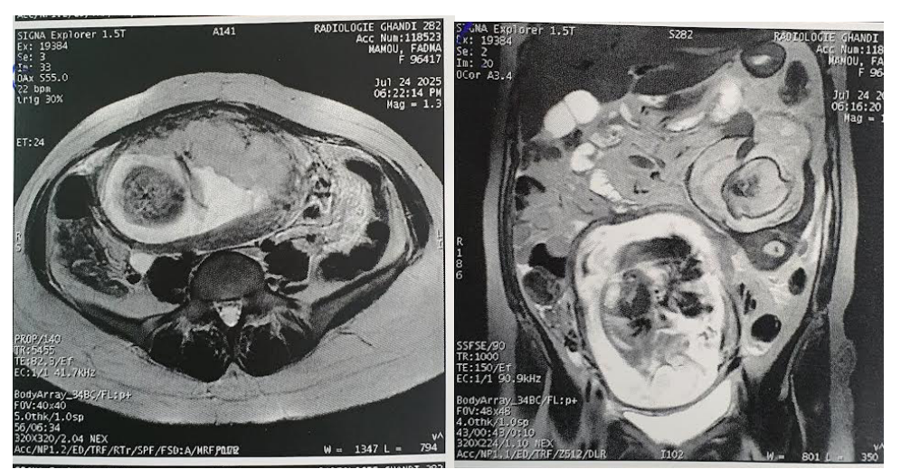
Figure 1 and 2: Abdominal MRI showed a uterine malformation with a triplet gestation in the larger horn and two in a rudimentary left horn, likely arrested, with a high risk of rupture.
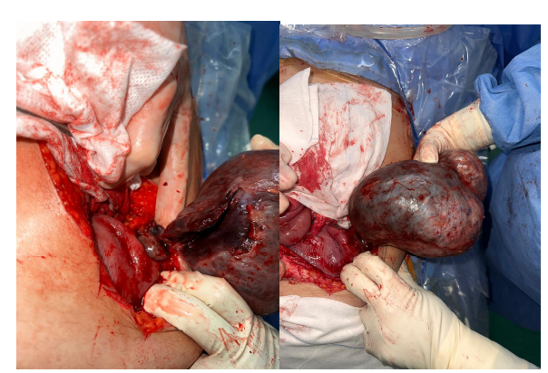
Figure 2.
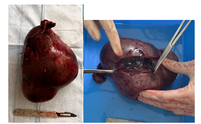
Figure 3. 10 Cm left ovarian mass with three turns of the pedicle.
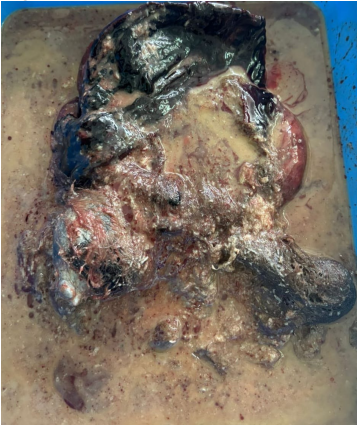
Figure 4. Ovarian teratoma.
Discussion
Teratomas encountered during pregnancy pose unique diagnostic and therapeutic challenges due to their rarity and overlapping presentation with common obstetric conditions.
Among these, mature ovarian teratomas—also known as dermoid cysts— are benign but may become symptomatic or complicated during gestation. In one recent case report, a 30-yearold pregnant woman presented with acute pelvic pain and a
large abdomino-pelvic mass. Imaging revealed a 15 × 15 cm
lesion, leading to surgical management via cystectomy. Histopathology confirmed a mature teratoma, and fertility was preserved through a conservative approach [2].
Mature teratomas are often asymptomatic, and many cases are
identified incidentally during routine prenatal ultrasound. Expectant management is feasible for small (< 6 cm), asymptomatic lesions, as they generally exhibit a favorable prognosis.
However, symptomatic or larger masses necessitate surgical
intervention, with timing and approach tailored to gestational
age and maternal–fetal risk [2].
In contrast, immature ovarian teratomas, though rare, present
greater concern. These malignant germ cell tumors require
prompt diagnosis and aggressive management. One report describes a pregnant patient diagnosed at 19 weeks with a left adnexal mass. The patient underwent salpingo-oophorectomy
and omental biopsy, revealing a Stage IA, Grade 2 immature
teratoma. Despite surveillance, metastatic disease emerged by
30 weeks. An early multidisciplinary intervention, including
timely chemotherapy, could potentially have mitigated progression. Ultimately, a cesarean section and oncologic resection were performed, followed by postoperative chemotherapy
using a BEP (bleomycin, etoposide, cisplatin) protocol. This
highlights both the aggressive behavior of high-grade immature teratomas and the importance of personalized, multidisciplinary care during pregnancy [3] [4].
Beyond ovarian presentations, fetal teratomas—such as sacrococcygeal teratomas (SCT)—carry significant fetal risk. Large
or malignant SCTs may lead to hydrops fetalis, high-output
cardiac failure, and fetal demise. One reported case involved a
fetal SCT detected at 16 weeks that expanded rapidly; the fetus unfortunately died in utero by 20 weeks, underscoring the
potential severity of such tumors and the importance of early
detection and monitoring [5].
Overall, several key principles emerge:
• Early detection and accurate classification (mature vs. immature; maternal vs. fetal) are vital. Imaging modalities like ultrasound and MRI, paired with tumor markers such as AFP and
LDH, can assist diagnosis, though interpretation must consider pregnancy-related physiological changes [3].
• Individualized, multidisciplinary management is essential.
For mature teratomas that are large or symptomatic, surgical
management—ideally with fertility preservation—is preferred. In contrast, high-grade immature teratomas may require early surgery combined with chemotherapy, even during
pregnancy, to improve outcomes [3,4].
• Maternal and fetal safety must be balanced. In cases such as
fetal SCTs, maternal monitoring, obstetric coordination, and
planning for immediate neonatal intervention are crucial if viable outcomes are possible [5].
• Finally, prognosis varies widely: mature ovarian teratomas
generally have excellent outcomes, while immature or fetal teratomas—with complications like metastatic spread, hydrops,
or mass effect—require closer surveillance and may carry
higher perinatal risk [2,3,5].
Conclusion
Teratomas in pregnancy are rare tumors that require timely diagnosis and individualized management.
While mature ovarian teratomas generally have favorable outcomes with conservative or surgical treatment, immature and
fetal forms carry significant risks and demand multidisciplinary care.
Early detection and close surveillance remain key to optimizing both maternal and fetal prognosis [2].
References
- Leiserowitz GS. Managing ovarian masses during pregnancy. Obstet Gynecol Surv. 2006;61(7):463-70. [PubMed.]
- Joudar et al, Mature teratoma during pregnancy: a case report. J Case Rep Images Obstet Gynecol. 2023. [Ref.]
- Vaidya et al, Immature Teratoma in Pregnancy: A Case Report. PMC. 2023. [Ref.]
- Hasdemir et al, Ovarian Immature Teratoma Detected During Pregnancy. Medical Science and Discovery. 2016. [Ref.]
- Tao et al, A fetal case of a large sacrococcygeal teratoma. Med Case Rep Study Protoc. 2022. [Ref.]