>Corresponding Author : Nouri M
>Article Type : Case Report
>Volume : 6 | Issue : 4
>Received Date : 22 August 2026
>Accepted Date : 1 September 2026
>Published Date : 3 September 2026
>DOI : https://doi.org/10.54289/JCRMH2600116
>Citation : Meriem N, Chahrazad E, Tawil FZ, Zakaria E, Bensouda MM, et al. (2026) Secondary Abdominal Pregnancy Arrested at 14 Weeks of Amenorrhea: a Case Managed at the Level III Maternity. J Case Rep Med Hist 6(4). doi: https://doi.org/10.54289 /JCRMH2600116
>Copyright : © 2026 Nouri M, et al. This is an open-access article distributed under the terms of the Creative Commons Attribution License, which permits unrestricted use, distribution, and reproduction in any medium, provided the original author and source are credited.
Case Report | Open Access | Full Text
Department of Gynecology and Obstetrics, Mother and Child Hospital, University Hospital Center (CHU) of Casablanca, Casablanca, Morocco
*Corresponding author: Nouri Meriem, Department of Gynecology and Obstetrics, Mother and Child Hospital, University Hospital Center (CHU) of Casablanca, Casablanca, Morocco
Abstract
Introduction: Abdominal pregnancy is a rare form of ectopic pregnancy, accounting for approximately 1% of cases. It carries a high risk of hemorrhage and maternal mortality.
Case Report: We report the case of a 28-year-old primigravida admitted with acute pelvic pain at 15 weeks + 2 days of
amenorrhea. Ultrasound revealed a 14-week fetus without cardiac activity, located in the left supra- and lateral-uterine region, associated with an abdominal placenta and hemoperitoneum. Surgical exploration confirmed a secondary abdominal
pregnancy complicated by left tubal rupture, with placental implantation on the omentum and sigmoid colon. A left salpingectomy was performed, with an uneventful postoperative recovery.
Conclusion: Abdominal pregnancy is a rare but serious condition. Diagnosis relies on imaging, and surgery remains the
treatment of choice. This case highlights the diagnostic challenge and the importance of prompt multidisciplinary management.
Keywords: Abdominal pregnancy, Ectopic pregnancy, Hemoperitoneum, Surgery, Case report
Introduction
Abdominal pregnancy is a rare and severe form of ectopic
pregnancy, defined by implantation of the gestational sac
within the peritoneal cavity, outside the uterus, fallopian
tubes, and ovaries. Its incidence is estimated between 1 in
10,000 and 1 in 30,000 pregnancies, representing about 1% of
ectopic pregnancies [1,2].
Despite its rarity, it constitutes a major cause of maternal morbidity and mortality, mainly due to massive intra-abdominal
hemorrhage related to placental implantation on highly vascularized structures [3,4]. Reported maternal mortality ranges
from 0.5% to 18%, while perinatal mortality frequently exceeds 80% [5].
Diagnosis is often difficult and delayed. Patients may present
with abdominal pain, vaginal bleeding, or acute abdomen. Ultrasound is the key diagnostic tool, but interpretation can be
hampered by associated uterine anomalies, and MRI may
sometimes be required to determine the exact site of placental
implantation [6,7].
Two forms are described: primary abdominal pregnancy,
which is extremely rare and defined by Studdiford’s criteria
[8], and secondary abdominal pregnancy, far more common,
resulting from tubal rupture or abortion with secondary implantation of the gestational sac in the peritoneal cavity.
We report a case of secondary abdominal pregnancy arrested
at 14 weeks of amenorrhea, complicated by hemoperitoneum
and associated with a polymyomatous uterus. This case, managed at the Level III Maternity Unit of Abderrahim
Harouchi Mother and Child Hospital (Casablanca University Hospital), illustrates the diagnostic and therapeutic challenges of this entity.
Case Report
We report the case of a 28-year-old primigravida, with no significant medical or surgical history, admitted to the Department of Gynecology and Obstetrics at the Level III Maternity Unit of Abderrahim Harouchi Mother and Child Hospital (Casablanca University Hospital) for acute pelvic pain
evolving for about 12 hours. Pregnancy was estimated at 15
weeks + 2 days of amenorrhea according to the last menstrual period .
Clinical Examination on Admission
On arrival, the patient was conscious (Glasgow score 15/15),
well oriented, and hemodynamically stable, although tachycardic: heart rate at 120 beats/min, blood pressure 140/90
mmHg, with a shock index of 0.8. Respiratory rate was 20 cycles/min, without signs of respiratory distress, and oxygen saturation was 96% in ambient air. Cardiopulmonary examination was normal.
Abdominal examination revealed moderate hypogastric
pain, without guarding or rebound tenderness, and the abdomen was soft. No external bleeding was noted.
Gynecological examination showed a long, closed, posterior
cervix, with no vaginal bleeding and no palpable lateral
uterine mass. Speculum examination revealed no blood at the
cervical os or in the vaginal cavity.
Complementary Investigations
Initial laboratory tests revealed:
Severe anemia with hemoglobin at 5.5 g/dl;
White blood cell count: 9,410/mm³;
Platelet count: 120,000/mm³;
Plasma β-hCG > 10,000 mIU/ml.
An urgent abdominal and pelvic ultrasound was performed,
showing: a polymyomatous uterus with irregular contours,
containing multiple heterogeneous hypoechoic masses, the
largest measuring 72 mm;
an endometrium that was difficult to identify;
an echogenic formation suggestive of placental tissue,
measuring 101 × 70 mm, with visible vascular pedicles;
a 14-week fetus located in the left supra- and lateral-uterine
region, without cardiac activity, with ultrasound signs of early fetal mummification (flattened skull, curved “pin-shaped”
spine); a moderate, finely echogenic peritoneal effusion, peri-uterine and extending to both flanks.
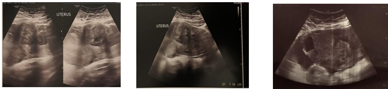
Figure 1. Ultrasound image showing the extra-uterine fetus in a left lateral and supra-uterine position.
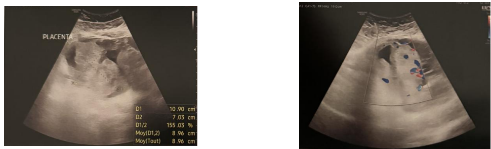
Figure 2. Ultrasound image showing the placental mass with vascular pedicles.
These findings suggested the diagnosis of an arrested abdominal pregnancy at 14 weeks of amenorrhea, complicated by
hemoperitoneum, in the context of a polymyomatous uterus.
Surgical Management
The patient was immediately hospitalized and stabilized (oxygen therapy, vascular filling, blood transfusion, and placement
of two large-bore peripheral intravenous lines). Given the presentation of hemoperitoneum, the decision was made to perform urgent surgical exploration. In the operating room, under
general anesthesia with rapid-sequence intubation, a Mouchel
incision was performed. Intraoperative findings revealed:
● a large amount of intra-abdominal effusion;
● an enlarged polymyomatous uterus;
● an extra-uterine intra-abdominal pregnancy with placental implantation on the omentum, the left iliac fossa, and the
sigmoid colon curvature;
● a hematoma adjacent to the meso-sigmoid, with no evidence of colonic perforation;
● an appendix that appeared macroscopically healthy,
though presenting with an apical hematoma;
● small bowel loops free of lesions.
The left fallopian tube was swollen and lacerated, requiring a
left salpingectomy.
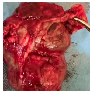
Figure 3. Surgical specimen showing the pathological left fallopian tube after salpingectomy, with trophoblastic tissue implantation.
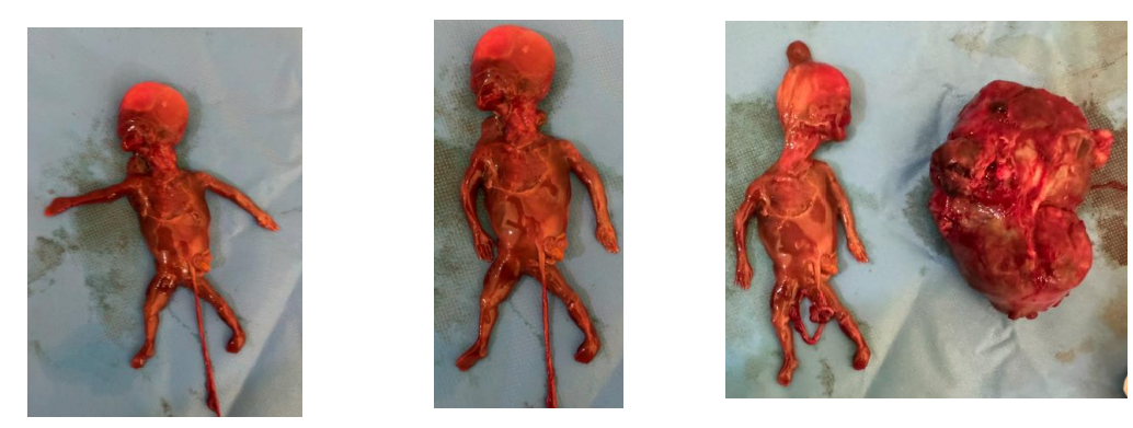
Figure 4. Surgical specimen showing the fetus extracted from the abdominal cavity.
An abundant lavage with normal saline was performed, followed by meticulous hemostasis. A Redon drain was placed
in the pouch of Douglas. The abdominal wall was closed layer
by layer after careful verification of all surgical materials.
The operative specimens (fetus, adnexa, placenta, and left fallopian tube) were sent for histopathological examination for confirmation.
Postoperative Course
The patient was transferred to the intensive care unit for close
postoperative monitoring. After 24 hours, she was transferred
to the postnatal ward. The postoperative course was favorable:
the drain output was approximately –50 ml on postoperative
day 2, after which it was removed. The patient was mobilized
early, with resumption of bowel function and progressive clinical improvement. She was discharged after hemodynamic stabilization and partial correction of anemia.
Discussion
Abdominal pregnancy is a rare but serious entity in obstetrics.
It accounts for about 1% of ectopic pregnancies, with a global incidence estimated between 1 in 10,000 and 1 in 30,000
pregnancies [1,2]. In low-resource countries, particularly in
sub-Saharan Africa and the Maghreb, the frequency is likely
underestimated due to delayed diagnosis and underreporting
[3].
In Morocco, ectopic pregnancies represent approximately 2 to
4% of gynecological emergencies [4], and abdominal localizations constitute a very small but particularly dangerous subset. Several studies conducted in Moroccan university hospitals, notably in Rabat, Fès, and Casablanca, report that abdominal pregnancies are often diagnosed in the second trimester,
sometimes even later, largely because of limited access to routine ultrasound in early pregnancy [5,6].
Maternal mortality associated with abdominal pregnancy remains high in developing countries, ranging from 3.5% to
18%, compared to less than 1% in high-income countries
[7,8]. Perinatal mortality frequently exceeds 80%, underscoring the severity of this condition [9]. These discrepancies reflect the decisive impact of socioeconomic factors and access
to healthcare on outcomes.
Clinically, the presentation is heterogeneous, ranging from
mild pelvic pain to acute hemorrhagic abdomen. In our case,
the patient presented with acute pelvic pain associated with severe anemia, consistent with the classical descriptions of delayed diagnosis in North Africa, where cases are often discovered during a complication [10].
Ultrasound is the key diagnostic tool, but its accuracy depends on several factors: quality of equipment, operator expertise, and the presence of associated uterine anomalies. In our
case, the polymyomatous uterus complicated interpretation.
Typical ultrasound findings include absence of myometrium
surrounding the pregnancy, abnormal localization of the fetus
in contact with digestive organs, and visualization of an extrauterine vascularized placenta [11]. In advanced centers, MRI
may be performed to determine placental topography and anticipate surgical difficulties [12].
From a physiopathological standpoint, two mechanisms are described: primary abdominal pregnancy, which is rare and
meets Studdiford’s criteria [13], and secondary abdominal
pregnancy, far more frequent, resulting from tubal rupture or
tubal abortion with secondary implantation of the gestational
sac. In our patient, intraoperative findings of a lacerated,
swollen left fallopian tube leave no doubt as to the secondary
nature of the pregnancy.
Placental implantation is a decisive factor for management
and prognosis. When the placenta is inserted into accessible
structures (parietal peritoneum, omentum), removal is generally feasible and safe. Conversely, implantation on richly vascularized organs (liver, mesentery, large vessels) may lead to catastrophic hemorrhage if excised, prompting some authors to recommend leaving the placenta in situ, combined with close
radiological and biological follow-up and sometimes adjuvant
methotrexate [14]. In our case, implantation on the omentum
and sigmoid colon curvature allowed relatively safe removal
during left salpingectomy.
Therapeutic management is primarily surgical. In early and
stable cases, laparoscopy may be considered. However, in
low-resource settings, laparotomy remains the standard, particularly in the presence of hemoperitoneum or advanced gestational age [15]. Our patient underwent laparotomy with a
Mouchel incision, a procedure adapted to the emergency, allowing complete exploration.
Maternal prognosis is strongly linked to the timeliness of diagnosis and availability of surgical and transfusion resources.
In Morocco and other African countries, diagnostic delays and
limited access to tertiary-level facilities continue to explain
significant maternal mortality [16]. Fetal prognosis is almost
uniformly poor, with mortality rates exceeding 80% and a
high risk of congenital malformations when abdominal pregnancies reach term [17].
Our case perfectly illustrates these challenges: a secondary abdominal pregnancy diagnosed in the second trimester, complicated by hemoperitoneum, but managed rapidly thanks to resources available at a university maternity hospital. It emphasizes the importance of early and systematic ultrasound, increased awareness among practitioners about this rare entity,
and multidisciplinary management to reduce maternal morbidity and mortality.
Conclusion
Although rare, abdominal pregnancy remains a formidable obstetric condition due to the severity of maternal complications
and the almost uniformly poor fetal prognosis. Diagnosis is often difficult and delayed, especially in low-resource settings
where access to systematic imaging is limited.
Our case illustrates a secondary abdominal pregnancy, complicated by hemoperitoneum, diagnosed in the second
trimester in the setting of a polymyomatous uterus, which contributed to diagnostic delay. Prompt surgical management,
combined with multidisciplinary monitoring at a tertiary-level
maternity unit, ensured a favorable maternal outcome. This
case highlights the importance of a high index of clinical suspicion, early high-quality ultrasound in pregnancy, and the
crucial role of referral centers in reducing maternal morbidity and mortality related to this exceptional condition.
References
- Nkusu Nunyalulendho D, Einterz EM. Advanced abdominal pregnancy: case report and review of 163 cases reported since 1946. Trop Med Int Health. 2008;13(7):943-946. [Ref.]
- Martin Jr JN, Sessums JK, Martin RW, Pryor JA, Morrison JC. Abdominal pregnancy: current concepts of management. Obstet Gynecol. 1988;71(4):549-57. [PubMed.]
- Baffoe P, Fofie C. Advanced abdominal pregnancy: the Ghanaian experience. J Obstet Gynaecol. 2011;31(1) :13-16. [Ref.]
- Benkaddour Y, Essatara Y, Asmouki H, Fichtali K, Aboulfalah A. Les grossesses extra-utérines au CHU de Rabat : étude de 450 cas. Pan Afr Med J. 2015;20:53. [Ref.]
- El Khouly NI, Fikri-Benbrahim K, Kharbach A, et al. Grossesses extra-utérines au Maroc : aspects épidémiologiques et thérapeutiques. East Mediterr Health J. 2013;19(3) :236-242. [Ref.]
- Filali Adib A, Moukit M, et al. Profil des grossesses extra-utérines au CHU de Casablanca. Rev Maroc Santé Reprod. 2018;5(2):25-32. [Ref.]
- Atrash HK, Friede A, Hogue CJ. Abdominal pregnancy in the United States: frequency and maternal mortality. Obstet Gynecol. 1987;69(3 Pt 1):333-7. [PubMed.]
- Costa SD, Presley J, Bastert G. Advanced abdominal pregnancy. Obstet Gynecol Surv. 1991;46(8):515-25. [PubMed.]
- Rahman MS, Al-Suleiman SA, Rahman J, Al-Sibai MH. Advanced abdominal pregnancy--observations in 10 cases. Obstet Gynecol. 1982;59(3):366-72. [PubMed.]
- Bouhouch N, et al. Grossesse abdominale secondaire à une rupture tubaire : à propos de 3 cas. Rev Maroc Gynécol Obstet. 2016;23(1):45-49. [Ref.]
- Onan MA, Turp AB, Saltik A, Akyurek N, Taskiran C, Himmetoglu O. Primary omental pregnancy: case report. Hum Reprod. 2005;20(3):807-9. [PubMed.]
- Worley KC, Hnat MD, Cunningham FG. Advanced extrauterine pregnancy: diagnostic and therapeutic challenges. Am J Obstet Gynecol. 2008;198(3):297.e1-297.e7. [Ref.]
- Studdiford WE. Primary peritoneal pregnancy. Am J Obstet Gynecol. 1942;44:487-491. [Ref.]
- Badria L, Amarin Z, Jaradat A, Zahawi H, Gharaibeh A, Zobi A. Full-term viable abdominal pregnancy: a case report and review. Arch Gynecol Obstet. 2003;268(4):340-2. [PubMed.]
- Varma R, Mascarenhas L, James D. Successful outcome of advanced abdominal pregnancy with exclusive omental insertion. Ultrasound Obstet Gynecol. 2003;21(2):192-4. [PubMed.]
- Traoré Y, et al. Grossesse abdominale avancée : aspects diagnostiques et thérapeutiques à Bamako. Mali Médical. 2017;32(4):18-24. [Ref.]