>Corresponding Author : Nouri M
>Article Type : Case Report
>Volume : 6 | Issue : 3
>Received Date : 22 August 2026
>Accepted Date : 1 September 2026
>Published Date : 3 September 2026
>DOI : https://doi.org/10.54289/JCRMH2600115
>Citation : Chaherazad E, Meriem N, Tawil FZ, Bensouda MM, Aicha G, et al. (2026) Ruptured Right Ampullary Tubal Ectopic Pregnancy with a Live Embryo Presenting as Hemoperitoneum in a Low-Risk Multigravida: a Case Report. J Case Rep Med Hist 6(3). doi: https://doi.org/10.54289/JCRMH2600115
>Copyright : © 2026 Etaouas Chaherazad, et al. This is an open-access article distributed under the terms of the Creative Commons Attribution License, which permits unrestricted use, distribution, and reproduction in any medium, provided the original author and source are credited.
Case Report | Open Access | Full Text
Department of Gynecology and Obstetrics, Mother and Child Hospital, University Hospital Center (CHU) of Casablanca, Casablanca, Morocco
*Corresponding author: Nouri Meriem, Department of Gynecology and Obstetrics, Mother and Child Hospital, University Hospital Center (CHU) of Casablanca, Casablanca, Morocco
Abstract
Background: Ectopic pregnancy (EP) complicates 1–2% of pregnancies and remains the leading cause of first-trimester
maternal mortality worldwide. Tubal rupture typically occurs early, and most ruptured pregnancies are no longer viable at
the time of presentation. We report a case of a ruptured ampullary tubal ectopic pregnancy in which the embryo remained
viable, presenting with hemoperitoneum at a gestational age beyond the usual window for tubal rupture.
Case presentation: A 28-year-old woman, gravida 2 para 1, with no notable medical, surgical, or gynecological history,
presented with acute pelvic pain, dark vaginal bleeding, and vomiting after two months of amenorrhea. She was hemodynamically stable with mild conjunctival pallor and right iliac fossa tenderness. Pelvic ultrasound showed a 10 × 6 cm
latero-uterine mass containing a gestational sac with an embryo corresponding to 12 weeks by crown–rump length and positive cardiac activity. Serum β-hCG exceeded 1395 mIU/mL. Surgical exploration confirmed a ruptured right ampullary
tubal pregnancy with moderate hemoperitoneum; a right salpingectomy was performed with peritoneal lavage and drain
placement. The post-operative course was uneventful, with stable hemoglobin and no transfusion requirement.
Conclusion: This case illustrates that tubal rupture with hemoperitoneum can occur in the ampullary segment beyond the
gestational age typically associated with rupture, even with a viable embryo and in the absence of identifiable risk factors.
It underscores the need for a high index of suspicion for ectopic pregnancy in any woman presenting with first-trimester
pain and bleeding, regardless of risk profile or reassuring hemodynamic status.
Keywords: Ectopic pregnancy, Tubal rupture, Hemoperitoneum, Ampullary pregnancy, Salpingectomy, Case report
Abbreviations: EP: Ectopic pregnancy
Introduction
Ectopic pregnancy (EP) — implantation of a blastocyst outside the uterine cavity — complicates approximately 1–2% of all pregnancies and remains the leading cause of pregnancy-related death in the first trimester worldwide [1,2]. Approximately 95–97% of ectopic pregnancies are tubal, and the ampullary segment is by far the most common implantation site, accounting for roughly 70% of cases [1]. Compared with the isthmic or interstitial segments, the ampulla is generally considered to carry a lower risk of rupture [3], and rupture classically presents early, often before 8 weeks of gestation, frequently in association with a non-viable pregnancy. Recognized risk factors include prior pelvic inflammatory disease, previous ectopic pregnancy, tubal surgery, smoking, intrauterine device use, and assisted reproduction [2,4,5]; however, most women who develop an ectopic pregnancy have no identifiable risk factor [2]. We report a case that is notable for two atypical features: tubal rupture occurred at a gestational age (12 weeks by crown–rump length) well beyond the usual window for ampullary rupture, and the embryo remained viable — with positive cardiac activity — at the time of rupture and surgery. We present this case in accordance with the CARE (CAse REport) guidelines [6,7].
Patient Information
A 28-year-old woman, gravida 2 para 1 (one prior living child, born by vaginal delivery), with no notable past medical, surgical, or gynecological history, was admitted to the emergency department for acute pelvic pain and dark (blackish) vaginal bleeding associated with vomiting, occurring in the context of a pregnancy dated to two months of amenorrhea.
Clinical Findings
On admission, the patient was conscious (Glasgow Coma Scale 15/15) and hemodynamically stable: blood pressure 110/70 mmHg, heart rate 80 bpm, oxygen saturation 100% on room air, respiratory rate 16/min, and temperature 36.7 °C. Conjunctivae were mildly pale. Urine output was preserved. Abdominal examination revealed a soft abdomen with tenderness in the right iliac fossa, without guarding or rebound tenderness. Gynecological examination (vulva and perineum) was unremarkable. Speculum examination showed a violaceous (bluish-discolored) cervix with clean vaginal walls.
Timeline
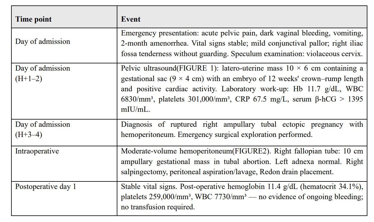
Table 1
Diagnostic Assessment
Pelvic ultrasound identified a latero-uterine (adnexal) mass
measuring 10 × 6 cm, containing a gestational sac of 9 × 4 cm
with an embryo whose crown–rump length corresponded to
12 weeks of gestation, with positive cardiac activity — a gestational age exceeding the two months estimated from the last
menstrual period, a discrepancy consistent with the diagnostic uncertainty inherent to ectopic pregnancies (Figure 1).
Laboratory investigations showed a hemoglobin of 11.7 g/dL,
a white cell count of 6830/ mm³, platelets of 301,000/mm³,
normal transaminases (AST 19 U/L, ALT 9 U/L), a Creactive
protein of 67.5 mg/L, and a serum β-hCG greater than 1395
mIU/mL — a value above the discriminatory zone at which an
intrauterine gestational sac would ordinarily be visualized by
transvaginal ultrasound in a normal pregnancy [8], supporting
the diagnosis of a pregnancy of extrauterine location. The combination of an empty uterine cavity, an adnexal gestational
mass with cardiac activity, and free intraperitoneal fluid is considered highly specific for ruptured ectopic pregnancy [9]. Together with the clinical picture — pelvic pain, dark vaginal
bleeding, a violaceous cervix, and mild pallor — these findings established the diagnosis of a ruptured right ampullary
tubal ectopic pregnancy complicated by hemoperitoneum.
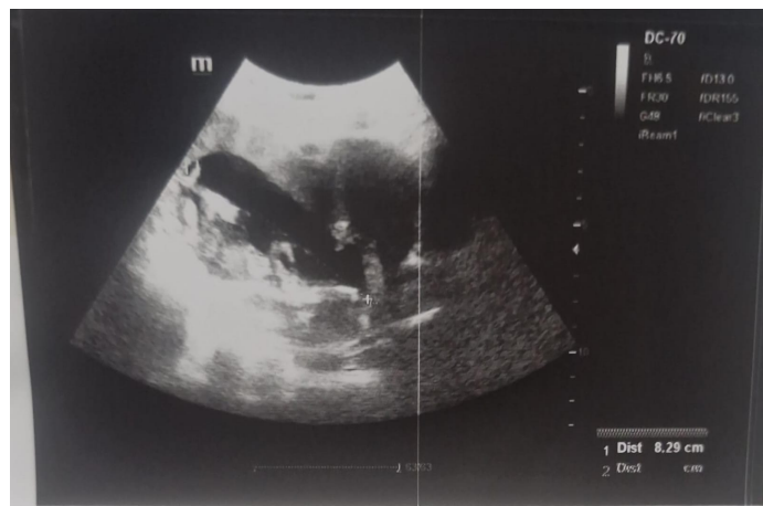
Figure 1. Latero-uterine mass 10 × 6 cm containing a kgestational sac (9 × 4 cm) with an embryo of 12 weeks.
Therapeutic Intervention
Given the sonographic evidence of tubal rupture with hemoperitoneum, the patient underwent emergency surgical exploration. This revealed a moderate-volume hemoperitoneum and a right fallopian tube harboring a 10 cm ampullary gestational mass — containing the gestational sac with positive cardiac activity described above — in a state of tubal abortion (Figure 2), (Figure 3). The left adnexa were normal. A right salpingectomy was performed, followed by aspiration and lavage of the peritoneal cavity and placement of a Redon drain (Figure 4).
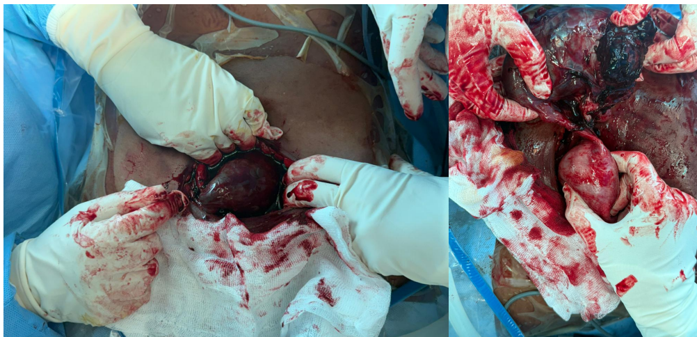
Figure 2. Moderate-volume hemoperitoneum.
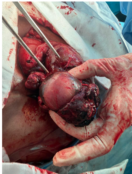
Figure 3. Right fallopian tube harboring a 10 cm ampullary gestational mass.
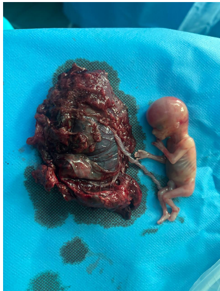
Figure 4. Gross specimen from the right salpingectomy, showing the ampullary segment of the fallopian tube distended by the gestational sac and hemorrhagic products of conception (left), along side the extracted embryo.
Follow-up and Outcome
The post-operative course was uneventful. Repeat blood count
on post-operative day 1 showed a hemoglobin of 11.4 g/dL
(hematocrit 34.1%, MCV 81.6 fL, MCHC 33.4 g/dL), a Intraoperative Moderate-volume hemoperitoneum((Figure 2).
Right fallopian tube: 10 cm ampullary gestational mass in
tubal abortion. Left adnexa normal. Right salpingectomy, peritoneal aspiration/lavage, Redon drain placement. Postoperative day 1 Stable vital signs. Post-operative hemoglobin 11.4
g/dL (hematocrit 34.1%), platelets 259,000/mm³, WBC
7730/mm³ — no evidence of ongoing bleeding; no transfusion
required. Time point Event white cell count of 7730/mm³, and
a platelet count of 259,000/mm³ — findings consistent with
hemodynamic stability and no evidence of ongoing intra-abdominal bleeding. No blood transfusion was required.
Laboratory Summary
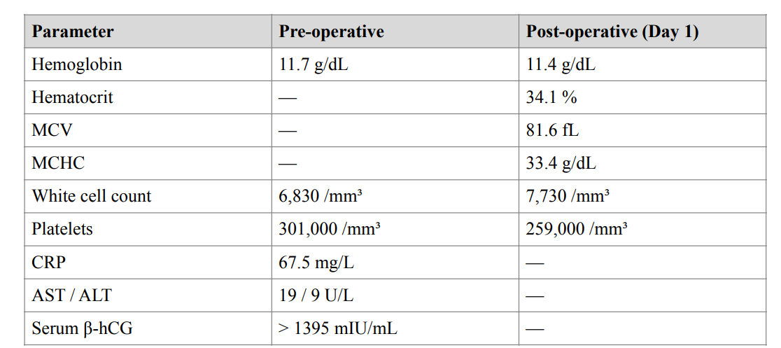
Table 2
Discussion
This case highlights two features that distinguish it from the
typical presentation of ampullary tubal rupture. First, rupture
occurred at a gestational age corresponding to 12 weeks by
crown–rump length, later than the window in which ampullary
pregnancies most commonly rupture; isthmic and interstitial
pregnancies are more classically associated with later, more
catastrophic rupture, whereas the ampulla — the site of roughly 70% of tubal pregnancies — is generally regarded as lower-risk and more likely to present earlier or to resolve as a
tubal abortion rather than a violent rupture [1,3]. Second, the
embryo remained viable, with positive cardiac activity documented on the pre-operative ultrasound performed shortly before surgery — an unusual finding, since ongoing hemorrhage
into the tube and peritoneal cavity is more often associated
with a non-viable gestation.
No conventional risk factor for ectopic pregnancy — prior
pelvic inflammatory disease, previous ectopic pregnancy,
tubal or pelvic surgery, intrauterine device use, smoking, or assisted reproduction — was identified in this patient, consistent
with the observation that a substantial proportion of women
who develop an ectopic pregnancy have no recognizable risk
factor [2]. This reinforces that a reassuring history cannot be
used to lower clinical suspicion when a woman presents with
first-trimester pain and bleeding.
Diagnostically, the discordance between the gestational age estimated from the last menstrual period (approximately 8 weeks) and the sonographic crown–rump length (12 weeks) illustrates a recognized difficulty in dating ectopic pregnancies,
and the elevated β-hCG value Parameter Pre-operative Post-
-operative (Day 1) Hemoglobin 11.7 g/dL 11.4 g/dL Hematocrit — 34.1 % MCV — 81.6 fL MCHC — 33.4 g/dL White
cell count 6,830 /mm³ 7,730 /mm³ Platelets 301,000 /mm³
259,000 /mm³ CRP 67.5 mg/L — AST / ALT 19 / 9 U/L —
Serum β-hCG > 1395 mIU/mL — in this case — above commonly cited discriminatory thresholds [8] — together with an
empty uterine cavity, appropriately raised suspicion for an extrauterine gestation. Diagnosis was corroborated by the combination of an adnexal mass with a live embryo and free peritoneal fluid, findings with high specificity for ruptured tubal
ectopic pregnancy on ultrasound [9].
Regarding management, salpingectomy — as performed in
this case — is generally recommended when the tube is extensively damaged or ruptured, when bleeding is not readily controlled, or when the ectopic mass is large (5 cm or greater), as
was the case here [1]. Randomized data comparing salpingectomy with the more conservative salpingostomy show similar
rates of subsequent intrauterine pregnancy and ectopic recurrence between the two approaches [1], supporting salpingectomy as an appropriate choice in the presence of rupture and a
large tubal mass, irrespective of future fertility considerations,
particularly when the contralateral tube is normal, as in this patient.
This case adds to the literature illustrating that the classical
teaching — that ampullary rupture occurs early and is associated with a non-viable pregnancy — does not preclude later, still-viable presentations. It reinforces the importance of early,
protocolized use of transvaginal ultrasound and serum β-hCG
in any woman of reproductive age presenting with pelvic pain
and abnormal bleeding, and of maintaining surgical readiness
even when initial hemodynamic parameters are reassuring.
Patient Perspective: Not available for this report.
Declarations: Conflicts of interest: none declared. Funding: none received.
This report was prepared in accordance with the CARE guidelines for case reports.
References
- Panelli DM, Phillips CH, Brady PC. Incidence, diagnosis and management of tubal and nontubal ectopic pregnancies: a review. Fertil Res Pract. 2015;1:15 [PubMed.]
- Flanagan HC, Duncan WC, Lin CJ, Spears N, Horne AW. Recent advances in the understanding of tubal ectopic pregnancy. Fac Rev. 2023;12:26. [PubMed.]
- Boykin T. Ipsilateral recurrent tubal ectopic pregnancy following a salpingectomy. J Diagn Med Sonography. 2016; 33(2):114-119. [PubMed.]
- Ectopic pregnancy. In: StatPearls [Internet]. Treasure Island (FL): StatPearls Publishing; 2025. [Ref.]
- Wang Y, Chen L, Tao Y, Luo M. Risk factors of ectopic pregnancy after in vitro fertilization-embryo transfer in Chinese population: A meta-analysis. PLoS One. 2024; 19(1):e0296497. [PubMed.]
- Gagnier JJ, Kienle G, Altman DG, Moher D, Sox H, Riley D. The CARE guidelines: consensus-based clinical case report guideline development. J Diet Suppl. 2013;10(4) :381-90. [PubMed.]
- Riley DS, Barber MS, Kienle GS, Aronson JK, von Schoen-Angerer T, Tugwell P, et al. CARE guidelines for case reports: explanation and elaboration document. J Clin Epidemiol. 2017;89:218-235. [PubMed.]
- Condous G, Kirk E, Lu C, Van Huffel S, Gevaert O, De Moor B, et al. Diagnostic accuracy of varying discriminatory zones for the prediction of ectopic pregnancy in women with a pregnancy of unknown location. Ultrasound Obstet Gynecol. 2005;26(7):770-5. [PubMed.]
- Ectopic pregnancy, ultrasound. In: StatPearls [Internet]. Treasure Island (FL): StatPearls Publishing; 2023. [Ref.]