>Corresponding Author : Nouri M
>Article Type : Case Report
>Volume : 6 | Issue : 2
>Received Date : 25 June, 2026
>Accepted Date : 07 July, 2026
>Published Date : 13 July, 2026
>DOI : https://doi.org/10.54289/JCRMH2600111
>Citation : Nouri M, Sahel I, Tawil F, Benchrifi Y, Benhassou M, et al. (2026) Neurofibromatosis Type 1 Associated with Breast Cancer: A Case Report. J Case Rep Med Hist 6(2): doi https://doi.org/10.54289/JCRMH2600111
>Copyright : © 2026 Nouri M, et al. This is an open-access article distributed under the terms of the Creative Commons Attribution License, which permits unrestricted use, distribution, and reproduction in any medium, provided the original author and source are credited.
Case Report | Open Access | Full Text
1Centre Mohammed VI for Cancer Treatment — CHU Ibn Rochd, Casablanca, Morocco
*Corresponding author: Nouri M, Centre Mohammed VI for Cancer Treatment — CHU Ibn Rochd, Casablanca, Morocco
Abstract
Neurofibromatosis type 1 (NF1) is an autosomal dominant neurocutaneous disorder caused by loss-of function mutations in the NF1 tumor suppressor gene encoding neurofibromin, a negative regulator of the RAS/MAPK signaling pathway. Women with NF1 carry a significantly elevated risk of breast cancer, particularly before the age of 50. We report the case of a 59-year-old woman with known NF1 who developed an invasive ductal carcinoma (IDC) of the left breast, not otherwise specified (NOS), classified cT4d N1c M1, of Luminal B molecular subtype (ER 60%, PR 10%, Ki67 20%, HER2 1+). The clinical presentation was dominated by massive cutaneous involvement with diffuse neurofibromas and an ulcerated, fungating left breast tumor with carcinomatous lymphangitis. Imaging revealed extensive NF1related lesions paravertebral plexiform neurofibromas, hepatic and renal cysts as well as severe cardiopulmonary complications that ultimately precluded surgical intervention. The patient received neoadjuvant chemotherapy (EC100 followed by Docetaxel), then endocrine therapy (Tamoxifen, subsequently switched to Letrozole + Palbociclib), with transition to integrated palliative care. This case highlights the complexity of breast cancer management in NF1 patients: diagnostic challenges posed by multiple cutaneous lesions, difficulties in radiological interpretation, elevated surgical risk, and the need for a comprehensive multidisciplinary approach.
Keywords: Neurofibromatosis Type 1; Breast Cancer; Invasive Ductal Carcinoma; Luminal B; Plexiform Neurofibroma; Palliative Care
Abbreviations: NF: Neurofibromatosis, IDC: Invasive Ductal Carcinoma, NOS: Not Otherwise Specified, GAP: GTPase-Activating Protein, MPNST: Malignant Peripheral Nerve Sheath Tumors, UOQ: Upper Outer Quadrant, NST: Nonspecific Type, SBR: Scarff-Bloom-Richardson, IHC: Immuno Histo Chemistry, ER: Estrogen Receptor, PR: Progesterone Receptor, IDC: Invasive Ductal Carcinoma, SIR: Standardized Incidence Ratio
Introduction
Neurofibromatosis type 1 (NF1), also known as von Recklinghausen disease, is an autosomal dominant genetic condition with an estimated incidence of 1 in 3,000 live births. It is caused by inactivating mutations in the NF1 tumor suppressor gene, located on chromosome 17q11.2, which encodes neurofibromin — a GTPase-activating protein (GAP) that negatively regulates RAS signaling through the MAPK/ERK pathway [1].
Classic clinical manifestations include café-au-lait macules, cutaneous and plexiform neurofibromas, axillary and inguinal freckling, Lisch nodules of the iris, and optic pathway gliomas. However, NF1 is also associated with a substantially increased risk of malignant tumors, including malignant peripheral nerve sheath tumors (MPNST), high-grade gliomas, gastrointestinal stromal tumors, and — of particular relevance — breast cancer [2].
Multiple epidemiological studies have established that women with NF1 carry a two- to fourfold increased risk of breast cancer compared to the general population, with a particularly pronounced excess risk before age 50. A cumulative lifetime breast cancer risk of 11–14% before age 50 has been estimated in NF1 women, compared to approximately 2.4% in the general population [3,4]. This risk profile is comparable to that associated with moderate-penetrance mutations in genes such as ATM or CHEK2, and NF1 is now recognized by some international guidelines as a genetic risk factor warranting enhanced breast cancer surveillance — including annual breast MRI from age 30–35 [5].
Despite this recognized association, breast cancer in the context of NF1 remains underreported in the literature, and its clinical management presents unique challenges that deserve detailed description. We present a clinical case illustrating the diagnostic and therapeutic difficulties encountered in this rare clinical setting.
Case Presentation
Patient History and Initial Presentation
A 70-year-old woman was admitted to Ibn Rochd University Hospital on December 31, 2024, presenting with a left breast mass that had been evolving for more than one month. Clinical examination revealed an inflammatory left breast with a large mass occupying nearly the entire breast. No palpable axillary lymphadenopathy was identified.
A 59-year-old woman, single, residing in Casablanca, with no tobacco, alcohol, or cannabis use, was under long-term follow-up in the dermatology department for previously diagnosed neurofibromatosis type 1. She reported no significant surgical history and no notable gyneco-obstetrical history. No family history of NF1 or breast cancer was documented.
The patient was referred to the Centre Mohammed VI for Cancer Treatment (CHU Ibn Rochd, Casablanca) for investigation of a left breast lesion that had been progressively evolving over the preceding 4 months. The chief complaints were:
Appearance of a painful left areolar nodule
Bloody nipple discharge
Physical Examination
General condition: ECOG Performance Status 1.
Dermatological examination: The patient presented with a striking and massive cutaneous involvement characteristic of severe NF1. Examination revealed hundreds of cutaneous neurofibromas of widely varying sizes, from a few millimetres to several centimetres in diameter, diffusely distributed across the entire trunk, back, bilateral upper and lower extremities, and abdomen (Figure 1). Several neurofibromas were pedunculated and particularly voluminous, notably at the lumbar and abdominal levels. The background skin demonstrated diffuse café-au-lait pigmentation.
Breast examination:
Left breast: Markedly enlarged, indurated, and erythematous, consistent with an inflammatory presentation. Palpation revealed a hard, fixed mass occupying the entirety of the left breast, with a large ulcerated, fungating areolar lesion exhibiting signs of secondary infection, without serous discharge. Nipple retraction was present (Figure 2).
Right breast: No palpable abnormality.
Axillary and supraclavicular lymph node regions: No clinically palpable lymphadenopathy.
Investigations
Breast Imaging
Mammography and breast ultrasound
Right breast:
Multiple bilateral hamartomatous formations.
Scaoered microcalcifications throughout the parenchyma.
No suspicious mass or architectural distortion (Figure 3).
Left breast:
Large cutaneous ulceration of the left breast (compression not tolerated by the patient for mammography).
On ultrasound: large ulceration of the left breast parenchyma; multiple bilateral hamartomatous formations (Figure 4).
Classification: ACR BIRADS 6 (left) — ACR BIRADS 4 (right)
Breast MRI:
Left breast:
Retroareolar large spiculated mass, hypointense on T1-weighted sequences, heterogeneously hyperintense on T2, with gadolinium enhancement demonstrating a type 2 enhancement curve, measuring 42 × 26 mm.
The mass emitted spicules extending to the pectoralis major muscle and was responsible for skin retraction with cutaneous thickening measuring up to 5 mm in maximal thickness.
Infracentimetric intra-mammary lymph node in the upper outer quadrant (UOQ), measuring 8 mm in short axis.
Axillary lymph nodes present, the largest measuring 9 mm in short axis.
Multiple polyploidal exophytic cutaneous lesions, hypointense on T1, hyperintense on T2, consistent with cutaneous neurofibromas (NF1 stigmata).
Right breast:
Oval, well-circumscribed lesions in the UOQ measuring 7 × 3 mm and 12 × 6 mm, consistent with intramammary lymph node.
Small cystic formation in the UOQ at the dermal level, measuring 5 mm in diameter.
Additional finding: Right-lateralized dorsal paravertebral cystic formation, in keeping with a dorsal cystic neurofibroma.
Overall MRI classification: ACR BIRADS 6 (left) — ACR BIRADS 2 (right)
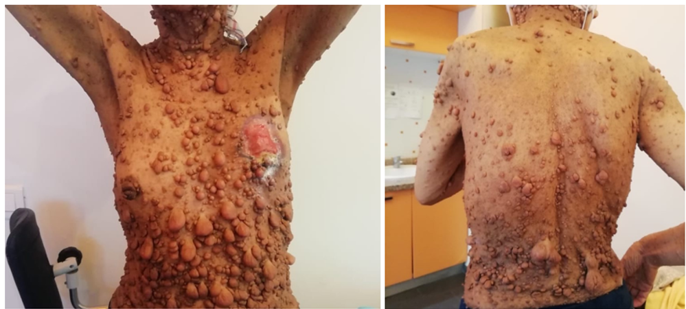
Figure 1. Cutaneous neurofibromas of widely varying sizes.
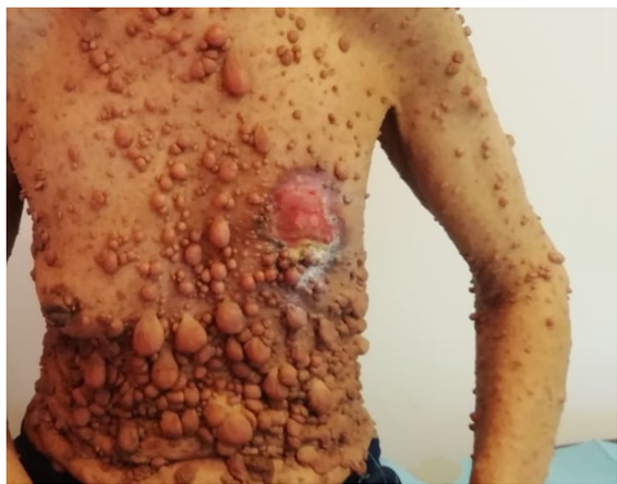
Figure 2. Left breast, with a large ulcerated.
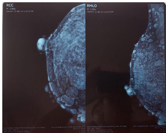
Figure 3. Mamography of right breast.
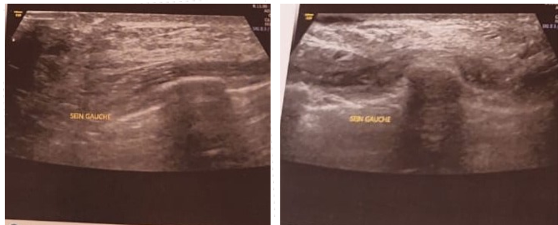
Figure 4. Ultrasound of the left breast parenchyma with a large ulceration.
Histopathology
Two biopsy fragments measuring 1 cm and 0.5 cm in greatest dimension were submitted.
Histopathological examination demonstrated cutaneous Tissue with dermis massively infiltrated by an invasive carcinomatous tumor proliferation arranged in cohesive clusters, solid nests, and occasional glandular formations. Tumor cells were of intermediate size with moderately atypical hyperchromatic nuclei, abundant eosinophilic cytoplasm, and a mitotic count of 14 mitoses/2 mm². There was an extensive carcinomatous lymphangitis with surface epidermal ulceration. The stromal reaction was moderate and predominantly lymphocytic. No perineural invasion was identified.
Pathological conclusion:
Carcinomatous lymphangitis with epidermal ulceration arising from an invasive breast carcinoma of nonspecific type (NST/NOS), Scarff-Bloom-Richardson (SBR) grade II (2+2+3).
Immunohistochemistry (IHC):
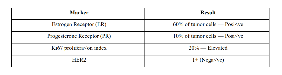
Molecular subtype: Luminal B, HER2-negative
(Note: HER2 testing was initially unavailable due to reagent stock shortage; subsequently confirmed as 1+ on repeat testing.)
Staging Workup
Thoraco-abdomino-pelvic CT scan:
Multiple hepatic nodular lesions, the largest measuring 19 × 12.7 mm at segment II, secondary origin initially not excluded.
Posterior paravertebral thoracic mass with endocanalicular extension, and left retroperitoneal mass, consistent with plexiform neurofibromas.
Scintigraphic correlation: hypofixation at the left 5th and 6th costospinal gutters, corresponding on CT to bone lysis in the vicinity of a parenchymal pulmonary mass.
Abdominal ultrasound and dedicated hepatic assessment:
Multiple rounded anechoic hepatic formations, non-vascularized on Doppler, without distinct walls — measuring 1.4 × 1 cm and 1.46 × 1.06 cm — consistent with simple benign hepatic cysts (segments II and VII).
Large right upper pole simple renal cyst.
Uterus with myomatous appearance.
Conclusion: hepatic lesions confirmed as cystic (benign), allowing reclassification from M1 to M0.
Spinal MRI:
Dorsal spinal neurofibroma at D6-D7-D8 with foraminal widening and extension to the left costovertebral groove in an hourglass configuration, causing moderate epidural compression.
Early degenerative lumbar spine changes.
Thoracic CT scan:
Left paravertebral well-defined formation at D7, enlarging the left neural foramen, with partially calcified wall, non-enhanced, measuring 54 × 45mm extending over 67mm in craniocaudal direction- consistent with a large plexiform neurofibroma in contact with the descending thoracic aorta.
Bilateral peri-hilar consolidations with air bronchogram.
Left pleural effusion of moderate abundance with ipsilateral mediastinal shift.
Pericardial effusion most pronounced at the left lateral recess, measuring 9 mm in maximal thickness.
Left supraclavicular lymph node measuring 7 mm in short axis.
Multiple hypodense hepatic lesions (cystic, previously characterized).
Right parapyelic renal cyst measuring 78 × 73 mm (Bosniak category I).
Multiple cutaneous nodules along the thoracic and abdominal wall consistent with known NF1 neurofibromas.
Thoracic CT scan:
Bilateral alveolar filling, ground-glass opacities in a “butterfly wing” pattern with air bronchogram.
Bilateral pleural and fissural effusion of moderate abundance, loculated on the right.
No pulmonary nodule or mass.
Cardiomegaly.
Conclusion: Acute pulmonary edema pattern, bilateral pleural effusion, cardiomegaly.
Brain MRI: Normal.
Cardiac Assessment
Echocardiography (pre-chemotherapy):
Non-dilated, non-hypertrophied left ventricle, preserved global and segmental contractility, LVEF 55‒60%.
Normal mitral inflow profile. Non-elevated LV filling pressures.
Non-dilated right ventricle with preserved longitudinal systolic function.
Minimal tricuspid regurgitation, estimated PASP 28 mmHg.
No pericardial effusion at this assessment.
Bronchoscopy:
Indication: mediastinal process with bilateral pleuropneumopathy in a patient with breast cancer and NF1.
Findings: Left bronchi with diffuse grade 3 inflammatory changes, mucosal thickening, and abundant whitish removable secretions. Right bronchi essentially normal.
Bronchoalveolar lavage (middle lobe): 240 cc instilled, 66 cc retrieved.
Staged bilateral bronchial biopsies performed.
Microbiological analyses: Pneumocystis jirovecii PCR, BK/Xpert, bacterial cultures, and cytological examination.
Transbronchial biopsies not performed due to risk of pneumothorax (patient with severe paroxysmal cough).
Final Diagnosis and Staging
Diagnosis: Invasive ductal carcinoma (IDC) of the left breast, not otherwise specified (NOS), Luminal B HER2-negative subtype.
TNM Classification:
Initial staging: cT4d N1c M1 (with Initial suspicion of hepatic and osseous metastases)
Revised staging after complete workup: cT4b N+ M0 (following confirmation of benign cystic hepatic lesions and NF1 origin of vertebral lesions)
Setting: Patient with known severe NF1, massive cutaneous neurofibroma involvement, paravertebral plexiform neurofibromas D6–D8 (hourglass configuration), cardiopulmonary complications (pleural effusion, pericardial effusion, recurrent inflammatory pneumonitis), and hepato-renal cystic disease.
Treatment
Neoadjuvant Chemotherapy
Multidisciplinary tumor board decision: Initiation of neoadjuvant chemotherapy with curaOve intent; staging of the case in the oncomedical RCP (Réunion de Concertation Pluridisciplinaire).
Phase 1 — Anthracycline-based regimen: EC100 x 3
Epirubicin 100 mg/m² + Cyclophosphamide
Tolerance: Good. No major toxicity reported.
Phase 2 — Taxane-based regimen: Docetaxel x3
Tolerance: ECOG PS 1, grade 1 mucositis, no significant toxicity.
Local wound care of the left breast ulceration was maintained throughout chemotherapy.
(Note: Between July and October, the patient was maintained on Tamoxifen 20 mg/day pending completion of the staging workup and resolution of logistical issues related to MRI availability).
Post-Chemotherapy Evaluation and Surgical Decision
Restaging CT scan: No evidence of distant metastatic disease.
Clinical examination: Highly locally advanced, ulcerated, fungating, infected left breast mass, no major clinical response to neoadjuvant chemotherapy.
Thoracic CT: Grade 3 inflammatory pneumonitis with moderate pleural and pericardial effusion.
Pre-anesthetic consultation (CPA) with the anesthesia-intensive care team:
The patient was deemed inoperable due to an unacceptably high risk of intraoperative death, related to:
1. Severe cardiopulmonary comorbidities (pericardial and pleural effusions, recurrent pneumonitis, cardiomegaly).
2. Paravertebral plexiform neurofibromas at D6–D8 in an hourglass configuration, placing the spinal cord and great vessels at risk.
3. Massive local tumor extension (T4b/T4d) rendering R0 resection technically unfeasible.
Palliative Systemic Therapy and Supportive Care
Decision: Formal contraindication to surgery and radiotherapy. Transition to palliative systemic therapy:
Endocrine therapy: Switch from Tamoxifen to Letrozole (aromatase inhibitor) given the Luminal B subtype.
Targeted therapy: Addition of Palbociclib (CDK4/6 inhibitor, Ibrance) to Letrozole, in line with the standard of care for Luminal B metastatic/locally advanced breast cancer. Integrated palliative care (soins palliatifs integres).
Monthly clinical monitoring: CA15-3 tumor marker + photographic documentation by a family caregiver.
Outcome
The clinical course following Initiation of systemic therapy was characterized by:
Temporary disease stabilization under endocrine therapy, with persistence of the locally advanced ulcerated breast lesion.
Recurrent cardiopulmonary complications, including bilateral pleural effusions and recurrent inflammatory pneumonitis, requiring repeated evaluations.
Sustained impossibility of locoregional treatment (surgery or radiotherapy) due to the combination of tumor burden, NF1-related structural lesions, and cardiovascular fragility.
Definitive transition to integrated palliative care with a focus on quality of life, pain management, and local wound care.
Discussion
NF1 as a Breast Cancer Predisposition Syndrome
The association between NF1 and breast cancer has gained increasing recognition over the past two decades. Neurofibromin functions as a tumor suppressor by accelerating the intrinsic GTPase activity of RAS, thereby promoting conversion of active GTP-bound RAS to inactive GDP-bound RAS. Loss-of function mutations in NF1 result in constitutive RAS/MAPK/ERK pathway activation, driving uncontrolled cellular proliferation and, in the mammary epithelium, oncogenic transformation [6,7].
The epidemiological evidence is now robust: a meta-analysis of prospective and retrospective cohorts estimated a standardized incidence ratio (SIR) of approximately 3.5 for breast cancer in NF1 women under 50, with a cumulative risk of 14% by age 50 versus 2.4% in the general population [4]. Importantly, this excess risk diminishes significantly after age 50, unlike hereditary breast cancer syndromes such as BRCA1/2, where elevated risk persists throughout life.
Several international expert groups and genetic counseling networks now recommend annual breast
MRI surveillance (complementing mammography) beginning at age 30–35 in women with confirmed NF1 [5]. Our patient, despite known NF1 follow-up in dermatology, had not undergone dedicated breast surveillance — likely contributing to the advanced stage at diagnosis.
Diagnostic Challenges in the Context of Massive Cutaneous Neurofibroma Burden
The diagnostic delay in our case reflects a fundamental challenge in NF1-associated breast cancer: the massive and diffuse cutaneous neurofibroma burden severely compromises clinical breast examination. The ulcerated areolar lesion was not immediately recognized as malignant, and the pathology request itself listed pyoderma gangrenosum, Schwannoma, MPNST, and cutaneous metastasis as differential diagnoses — reflecting genuine clinical uncertainty.
This illustrates a critical point: in NF1 patients, any new or rapidly evolving cutaneous or subcutaneous lesion — particularly in the breast region — should prompt early histopathological evaluation rather than attribution to the underlying NF1 phenotype. The availability of a dedicated dermatologist and oncologist collaborating closely is essential.
Furthermore, physical examination alone is insufficient for breast surveillance in this population. Given the multiple cutaneous artifacts and the density of lesions, breast MRI is the modality of choice for surveillance and characterization, with ultrasound as a complementary tool.
Pitfalls in Radiological Interpretation
Radiological interpretation in NF1 patients is particularly challenging, as NF1-specific lesions may closely mimic malignant findings across multiple organ systems:
Paravertebral plexiform neurofibromas (hourglass configuration at D6-D8 in our case) can simulate spinal metastases, infiltrative lymphoma, or primary bone tumors on CT/MRI.
Hepatic cysts associated with NF1 were initially misinterpreted as potential hepatic metastases on CT, leading to an Initial M1 Classification that was subsequently corrected after dedicated MRI and ultrasound.
Bilateral breast hamartomatous formations, visible on both mammography and MRI, represent benign NF1-specific lesions that may mask underlying malignant foci or generate false-positive findings.
Cutaneous neurofibromas themselves produce multiple skin nodules visible on all imaging modalities, requiring careful correlation with clinical findings.
In our case, the Initial overstaging (M1 disease) significantly influenced the therapeutic strategy and nearly led to a purely palliative approach without neoadjuvant chemotherapy. Careful multidisciplinary radiological review with NF1 expertise is therefore essential before Definitive staging decisions.
Molecular Subtype and Therapeutic Implications
The Luminal B HER2-negative subtype observed in our patient is the most commonly reported molecular subtype in NF1-associated breast cancers [8]. This subtype is characterized by hormone receptor positivity, elevated Ki67 (≥20%), and HER2 negativity — implying a biology of intermediate aggressiveness with proliferative features exceeding those of Luminal A tumors.
From a mechanistic standpoint, the constitutive activation of the RAS/MAPK/ERK pathway secondary to neurofibromin loss may contribute to the elevated proliferation index and potentially to endocrine resistance. Importantly, this molecular landscape creates several therapeutic opportunities:
CDK4/6 inhibitors (palbociclib, ribociclib, abemaciclib) combined with aromatase inhibitors represent the standard of care for HR+/HER2- advanced breast cancer and were appropriately incorporated in our patient’s treatment plan. Their mechanism of action — blocking RB phosphorylation and arresting cell cycle progression — acts downstream of the RAS/MAPK pathway, theoretically complementing neurofibromin loss.
MEK inhibitors (trametinib, cobimetinib) have demonstrated activity in NF1-associated MPNSTs and other NF1-driven malignancies; their potential in NF1-associated breast cancer warrants further investigation [9].
mTOR inhibitors (everolimus) may also be relevant given the frequent co-activation of the PI3K/AKT/mTOR pathway in RAS-driven tumors.
Surgical Risk and Anesthetic Considerations in NF1
The surgical contraindication in our patient was absolute and driven by a convergence of NF1-specific and oncological factors:
NF1-specific surgical risks:
The paravertebral plexiform neurofibroma at D6-D8 in an hourglass configuration creates a risk of intraoperative hemorrhage, aortic injury (given proximity to the descending thoracic aorta), and spinal cord damage with irreversible neurological sequelae.
General anesthesia in NF1 patients carries specific risks including airway neurofibromas, altered responses to neuromuscular blocking agents, and cardiovascular instability due to associated vasculopathy.
Oncological factors:
Massive T4d/T4b local extension with carcinomatous lymphangitis rendered R0 mastectomy technically unfeasible.
The inflammatory breast carcinoma phenotype (T4d) is itself associated with a high risk of local recurrence even after optimal surgery.
Cardiopulmonary factors:
Recurrent bilateral pleural and pericardial effusions, along with grade 3 inflammatory pneumonitis, created an unacceptable perioperative risk of cardiorespiratory decompensation.
This clinical scenario underscores the necessity of early pre-anesthetic consultation in all NF1 patients with breast cancer considered for locoregional treatment and highlights that standard oncological decision algorithms may require substantial modification in this population.
Conclusion
We report a rare and clinically complex case of neurofibromatosis type 1 associated with an advanced
Luminal B invasive ductal carcinoma of the left breast in a 59-year-old woman with massive cutaneous neurofibroma burden. This case contributes to the growing body of literature documenting the breast cancer predisposition conferred by NF1 and illustrates several critical clinical lessons:
1. Systematic breast MRI surveillance should be offered to all women with NF1 from age 30–35, in line with emerging international recommendations. Reliance on clinical examination alone is insufficient given the masking effect of cutaneous and subcutaneous neurofibromas.
2. Any new or evolving breast lesion in an NF1 patient — regardless of its clinical appearance — warrants prompt histopathological evaluation and should not be dismissed as a neurofibroma complication.
3. Radiological interpretation in NF1 requires specific expertise: paravertebral neurofibromas, hepatic and renal cysts, and hamartomatous breast formations can all closely mimic metastatic disease, potentially leading to overstaging and inappropriate therapeutic decisions.
4. Surgical planning in NF1 patients with breast cancer must integrate the specific anesthetic and structural risks related to plexiform neurofibromas and associated cardiovascular comorbidities. Early pre-anesthetic consultation is mandatory.
5. Targeted therapies — particularly CDK4/6 inhibitors and, in the future, MEK inhibitors — hold promise as systemic alternatives when locoregional treatment is contraindicated, and deserve dedicated investigation in the NF1-associated breast cancer population.
A multidisciplinary approach integrating dermatology, genetic counseling, medical oncology, surgery, interventional radiology, cardiology, and palliative care is essential to optimize outcomes and quality of life in this challenging clinical setting.
Conflict of interest: None declared.
Patient consent: Informed consent for publication of this case report, including clinical photographs and imaging data, was obtained in accordance with the institutional ethics committee requirements and the Declaration of Helsinki. All identifying information has been removed.
Acknowledgements: The authors thank the multidisciplinary team of the Centre Mohammed VI for Cancer Treatment, CHU Ibn Rochd, Casablanca, for their collegial contribution to this patient’s care.
References
- Legius E, Messiaen L, Wolkenstein P, et al. Revised diagnostic criteria for neurofibromatosis type 1 and Legius syndrome: an international consensus recommendation. Genet Med. 2021;23(8):1506–1513. [PubMed.]
- Evans DG, Howard E, Giblin C, et al. Birth incidence and prevalence of tumor-prone syndromes: estimates from a UK family genetic register service. Am J Med Genet A. 2010;152A(2):327–332. [PubMed.]
- Walker L, Thompson D, Easton D, et al. A prospective study of neurofibromatosis type 1 cancer incidence in the UK. Br J Cancer. 2006;95(2):233–238. [PubMed.]
- Madanikia SA, Bergner AL, Ye X, Blakeleft JO. Increased risk of breast cancer in women with NF1. Am J Med Genet A. 2012;158A(12):3056–3060. [Ref.]
- Villani A, Greer MLC, Kalish JM, et al. Recommendations for cancer surveillance in individuals with RASopathies and other rare genetic syndromes with increased cancer risk. Clin Cancer Res. 2017;23(12):e83–e90. [PubMed.]
- Cichowski K, Jacks T. NF1 tumor suppressor gene function: narrowing the GAP. Cell. 2001;104(4):593–604. [PubMed.]
- Johannessen CM, Reczek EE, James MF, Brems H, Legius E, et al. The NF1 tumor suppressor critically regulates TSC2 and mTOR. Proc Natl Acad Sci USA. 2005;102(24):8573–8578. [PubMed.]
- Wang X, Levin AM, Smolinski SE, Vigneau FD, Levin NK, et al. Breast cancer and other neoplasms in women with neurofibromatosis type 1: a retrospective review of cases in the Detroit metropolitan area. Am J Med Genet A. 2012;158A(12):3061–3064. [PubMed.]
- Blakeleft JO, Ye X, Duda DG, et al. Efficacy and biomarker study of bevacizumab for adults with neurofibromatosis type 1 associated with progressive plexiform neurofibromas. J Clin Cancer Res. 2016;22(21):5327–5335. [Ref.]