>Corresponding Author : Intissar Selmani
>Article Type : Case Report
>Volume : 6 | Issue : 3
>Received Date : 11 June, 2026
>Accepted Date : 25 June, 2026
>Published Date : 24 July, 2026
>DOI : https://doi.org/10.54289/JCRMH2600110
>Citation : Selmani I, Chaimaa EMED, Saligane A, Bencherifi Y, and Benhessou M. (2026) Encapsulated Papillary Carcinoma of the Breast with Minimal Invasive Component and Axillary Lymph Node Metastasis: A Case Report and Review of the Literature. J Case Rep Med Hist 6(3): doi https://doi.org/10.54289/JCRMH2600110
>Copyright : © 2026 Selmani I, et al. This is an open-access article distributed under the terms of the Creative Commons Attribution License, which permits unrestricted use, distribution, and reproduction in any medium, provided the original author and source are credited.
Case Report | Open Access | Full Text
1Department of Gynecology and Obstetrics, Ibn Rochd University Hospital Center, Casablanca, Morocco
2Department of Gynecology and Obstetrics, Ibn Rochd University Hospital Center, Casablanca, Morocco
*Corresponding author: Intissar Selmani, Department of Gynecology and Obstetrics, Ibn Rochd University Hospital Center, Casablanca, Morocco
Abstract
Background: Encapsulated papillary carcinoma (EPC) is a rare breast neoplasm accounting for less than 2% of all breast cancers. Although generally associated with an excellent prognosis, EPC may occasionally present with invasive carcinoma and lymph node metastasis.
Case Presentation: A 59-year-old postmenopausal woman presented with a progressively enlarging right breast mass. Mammography and ultrasonography revealed a suspicious cystic lesion classified as BIRADS 5. Core needle biopsy demonstrated an encapsulated papillary carcinoma of luminal B subtype. Breast-conserving surgery with sentinel lymph node biopsy followed by axillary lymph node dissection was performed. Histopathological examination revealed EPC associated with a 2-mm invasive carcinoma, high-grade ductal carcinoma in situ (DCIS), lymphovascular invasion, and nodal involvement.
Conclusion: This case highlights the importance of extensive histological sampling and careful assessment of extracapsular invasion in EPC, particularly when adverse prognostic factors are present.
Keywords: Encapsulated Papillary Carcinoma; Breast Cancer; Papillary Lesion; Invasive Carcinoma; Lymphovascular Invasion; Lymph Node Metastasis
Abbreviations: EPC: Encapsulated Papillary Carcinoma, DCIS: Ductal Carcinoma in Situ, CC: Craniocaudal, MLO: Mediolateral Oblique
Introduction
Encapsulated papillary carcinoma (EPC) is an uncommon papillary breast neoplasm characterized by papillary proliferations enclosed within a thick fibrous capsule and typically lacking a peripheral myoepithelial cell layer [1-3]. It predominantly affects postmenopausal women and usually exhibits an indolent clinical course [1,2]. However, EPC may be associated with ductal carcinoma in situ (DCIS), invasive carcinoma, and, rarely, lymph node metastasis [4,5]. Recent studies have highlighted the importance of identifying invasive foci because they influence staging, prognosis, and treatment strategies [1,4,6].
We report a case of EPC associated with a minimal invasive component and axillary nodal involvement and review the current literature.
Case Presentation
A 59-year-old postmenopausal woman (G3P3) presented with a progressively enlarging right breast mass that had evolved over two months. She had no personal history of malignancy and no family history of breast cancer.
Clinical examination revealed a freely mobile, well-circumscribed mass measuring approximately 2 cm at the junction of the inner quadrants of the right breast. No palpable axillary lymphadenopathy was detected.
Mammography demonstrated a well-defined oval high-density mass located in the lower inner quadrant of the right breast (Figure 1A and 1B).
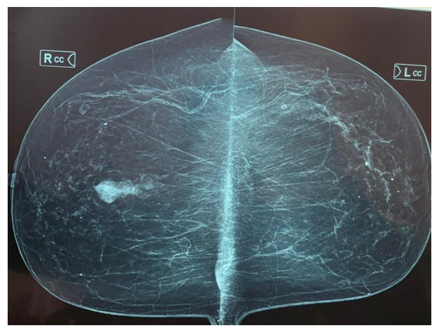
Figure 1A. Bilateral mammography, craniocaudal (CC) view showing a well-circumscribed mass in the lower inner quadrant of the right breast.
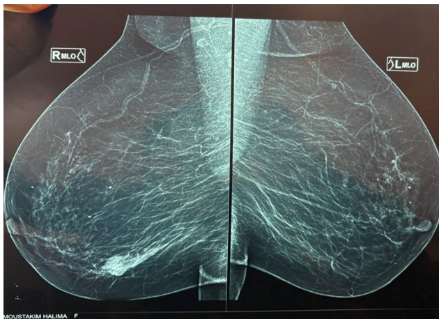
Figure 1B. Bilateral mammography, mediolateral oblique (MLO) view demonstrating the same lesion.
Breast ultrasonography revealed a complex cystic lesion with intracystic echogenic content and associated small satellite cystic lesions (Figure 2). The lesion was categorized as BIRADS 5. A right axillary lymph node measuring 7 mm was also identified.
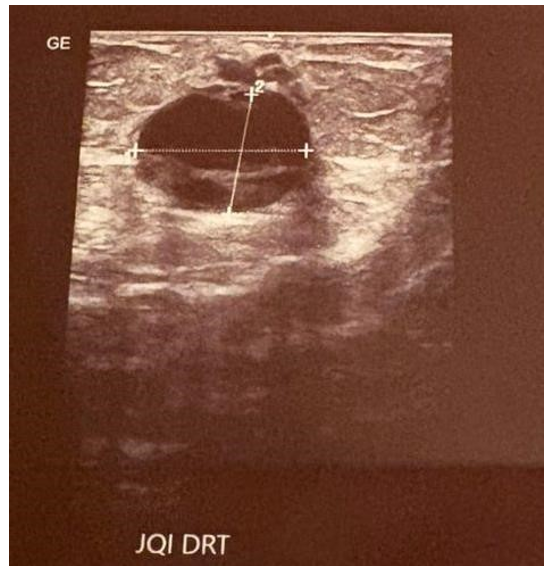
Figure 2. Breast ultrasound showing a complex cystic lesion with intracystic solid components corresponding to the encapsulated papillary carcinoma.
Ultrasound-guided core needle biopsy demonstrated encapsulated papillary carcinoma with an invasive component. Immunohistochemical analysis revealed strong estrogen receptor positivity (100%), absence of progesterone receptor expression (0%), HER2 negativity, and a Ki-67 proliferation index of 40%, consistent with a luminal B molecular subtype.
The patient underwent breast-conserving surgery with sentinel lymph node biopsy. Because the sentinel lymph node was positive for metastatic involvement, completion axillary lymph node dissection was subsequently performed.
Gross examination of the lumpectomy specimen revealed an oriented breast tissue specimen weighing 73 g and measuring 7 × 6.5 × 5.1 cm. Serial sectioning identified a poorly circumscribed, partially cystic lesion containing hemorrhagic material and measuring 5 × 3.5 × 2.2 cm.
Microscopic examination demonstrated an encapsulated papillary neoplasm surrounded by a thick fibrous capsule. The papillary structures were composed of delicate fibrovascular cores lined by atypical pseudostratified columnar epithelial cells exhibiting moderate to marked nuclear atypia and increased mitotic activity.
A focal invasive carcinoma measuring 2 mm was identified beyond the fibrous capsule in one of eighteen examined tissue blocks. The invasive component consisted of cords, nests, and glandular structures composed of atypical epithelial cells. Lymphovascular invasion was present.
An associated high-grade cribriform ductal carcinoma in situ with comedonecrosis represented approximately 10% of the lesion. Adjacent breast tissue exhibited fibrotic changes.
The final diagnosis was encapsulated papillary carcinoma associated with invasive carcinoma (pT1a) and lymphovascular invasion.
The patient was subsequently referred to the oncology department for adjuvant treatment.
Discussion
EPC represents a rare subtype of papillary breast carcinoma that predominantly affects elderly women [1,2]. Imaging findings are frequently nonspecific, often demonstrating a circumscribed cystic-solid lesion on mammography and ultrasonography, as observed in our patient [7,8].
Histologically, EPC is characterized by papillary proliferations enclosed within a fibrous capsule and the absence of myoepithelial cells at the periphery [2,5,6,9]. Immunohistochemical markers such as p63, calponin, and smooth muscle myosin heavy chain are useful in distinguishing EPC from benign papillary lesions [2,6].
The major challenge in EPC is the identification of invasive disease. According to Rakha et al., true invasion should be defined by neoplastic extension beyond the fibrous capsule into the surrounding stroma [5]. In our case, a 2-mm invasive focus was identified beyond the capsule, fulfilling the criteria for invasive carcinoma associated with EPC.
Most EPCs exhibit a luminal phenotype with strong hormone receptor expression and HER2 negativity [1,4]. However, our patient demonstrated a luminal B profile because of the elevated Ki-67 index and absence of progesterone receptor expression, suggesting a potentially more aggressive biological behavior.
The presence of lymphovascular invasion and nodal metastasis despite the minimal invasive component is noteworthy. Although uncommon, axillary lymph node involvement has been described in EPC associated with invasive carcinoma and supports the need for sentinel lymph node evaluation in selected cases [1,4,8].
Current management relies on complete surgical excision with negative margins [6,8]. Breastconserving surgery followed by radiotherapy is generally considered appropriate [8]. Endocrine therapy is recommended for hormone receptor-positive tumors, while chemotherapy may be considered in the presence of nodal involvement or other adverse prognostic features [1,4,8].
Conclusion
Encapsulated papillary carcinoma is an uncommon breast neoplasm with generally favorable prognosis. Nevertheless, associated invasive carcinoma, lymphovascular invasion, and lymph node metastasis may occur even when the invasive component is extremely limited. This case underscores the importance of meticulous pathological examination and multidisciplinary management to ensure accurate staging and optimal treatment.
References
- Tan HJ, Tan PH, Leong LCH, et al. Encapsulated papillary carcinoma of the breast: An institutional case series and literature review. Cancer Med. 2023;12(10):11408‒11416. [PubMed.]
- Ni Y, Tse GM. Papillary lesions of the breast ‒ review and practical issues. Semin Diagn Pathol. 2022;39(5):344‒354. [PubMed.]
- Mulligan AM. Encapsulated Papillary Carcinoma of the Breast. Surg Pathol Clin. 2009;2(2):319‒350. [PubMed.]
- Changrong W, Zhibo Z, Jufeng G, et al. Encapsulated Papillary Carcinoma of the Breast: A Review of Clinicopathologic Characteristics, Molecular Mechanisms, and Patient Management. Cancer Control. 2024;31:10732748241299071. [PubMed.]
- Rakha EA, Tun M, Junainah E, Ellis IO, Green A. Encapsulated papillary carcinoma of the breast: a study of invasion associated markers. J Clin Pathol. 2012;65(8):710‒714. [PubMed.]
- Kulka J, Madaras L, Floris G, Lax SF. Papillary lesions of the breast. Virchows Arch. 2022;480(1):65‒84. [PubMed.]
- Tse GM, Tan PH, Moriya T. The role of immunohistochemistry in the differential diagnosis of papillary lesions of the breast. J Clin Pathol. 2009;62(5):407‒413. [PubMed.]
- Athanasiou A, Khomsi F, de Joliniere B, Feki A. Encapsulated Papillary Carcinoma: A Case Report and Review of the Literature. Front Surg. 2022;8:743881. [PubMed.]
- Collins LC, Schnitt SJ. Papillary lesions of the breast: selected diagnostic and management issues. Histopathology. 2008;52(1):20‒29. [PubMed.]