>Corresponding Author : Bahlioui Fatima Ezzahra
>Article Type : Case Report
>Volume : 5 | Issue : 12
>Received Date : 26 Oct, 2025
>Accepted Date : 08 Nov, 2025
>Published Date : 03 Dec, 2025
>DOI : https://doi.org/10.54289/JCRMH2500160
>Citation : Ezzahra BF, Zahra CF, Benchrifi Y, Mustapha B, Mohamed E, et al. (2025) Uterine Sarcoma: 1 Case Report and Review of the Literature. J Case Rep Med Hist 5(12): doi https://doi.org/10.54289/JCRMH2500160
>Copyright : © 2025 Ezzahra BF, et al. This is an open-access article distributed under the terms of the Creative Commons Attribution License, which permits unrestricted use, distribution, and reproduction in any medium, provided the original author and source are credited.
Case Report | Open Access | Full Text
1Resident Physician, Department of Gynecology and Obstetrics at the Ibn Rochd University Hospital, Casablanca, Morocco
2Professor in the Department of Gynecology and Obstetrics at the Ibn Rochd University of Hospital in Casablanca Morocco
*Corresponding author: Bahlioui Fatima Ezzahra, Resident Physician, Department of Gynecology and Obstetrics, at Ibno Rochd University Hospital, Casablanca, Morocco
Abstract
Uterine sarcoma is a rare malignant tumor accounting for 2 to 5% of uterine cancers [1], with an annual incidence of 0.36 to 0.64 cases per 100,000 women [2]. The main subtypes are leiomyosarcoma, endometrial stromal sarcoma, undifferentiated sarcoma, adenosarcoma, and carcinosarcoma [3]. Symptoms are often nonspecific, such as abnormal uterine bleeding and pelvic pain [4]. Diagnosis is based on imaging and, above all, histological analysis [5].
Surgery, in the form of total hysterectomy with bilateral adnexectomy, is the standard treatment [6]. Depending on the histological type and stage, adjuvant treatments (radiotherapy, chemotherapy, or hormone therapy) may be offered [7,8]. The prognosis varies greatly: it is favorable for low-grade endometrial stromal sarcomas [9] but poor for undifferentiated forms or metastatic leiomyo sarcomas [10,11].
Improvements in survival are based on early diagnosis, individualized treatments, and the development of innovative therapies [12,13].
Keywords: Uterine Sarcoma, Leiomyo Sarcoma, Endometrial Stromal Sarcoma, Carcino Sarcoma, Rare Malignant Tumor, Hysterectomy, Prognosis
Introduction
Uterine sarcoma is a rare malignant tumor of the uterine body, originating from the myometrium or supporting connective tissue. It accounts for approximately 2 to 5% of uterine cancers [1] with an estimated incidence of between 0.36 and 0.64 cases per 100,000 women per year [2]. The main histological sub types are leiomyo sarcoma, endometrial stromal sarcoma, undifferentiated sarcoma, adenosarcoma, and carcinosarcoma [3]. Leiomyosarcoma is the most common and derives from uterine smooth muscle cells [14]. Endometrial stromal sarcoma can below-grade, with a good prognosis, or high-grade, which is more aggressive [15]. Carcino sarcoma, which accounts for up to 50% of uterine sarcomas, has a dual epithelial and mesenchymal component and is often treated as endometrial carcinoma [16]. Symptoms are often non specific: abnormal uterine bleeding, pelvic pain, or abdominal mass [4]. Diagnosis is based on imaging and, above all, histological examination after hysterectomy [5].
Case report
This is a 65-year-old female patient, mother of one child delivered vaginally, menopausal for 20 years, being treated for type 2 diabetes with oral anti diabetic medication, presenting with abnormal uterine bleeding that has been ongoing for 3 months with no other associated symptoms.
Clinical examination revealed a friable 7 cm mass protruding from the cervix that did not infiltrate the vaginal walls.
Pelvic MRI:en larged uterus with an endocavitary mass occupying almost the entire uterus, measuring 12x7x6cm. This mass appears to exceed 50% of the thickness of the myometrium, however there is a continuous external band of myometrium that has not been penetrated. At the bottom, it extends beyond the cervix at the level of the anterior wall in the upper third of the vagina. There is no evidence of invasion of the bladder or rectum, with no visible pelvic lymphadenopathy of significant size (Figure 1).
-A biopsy of the mass delivered through the cervix was performed, confirming a sarcomatoid tumor-like process.
-A total hysterectomy without preservation of the adnexa was performed, as well as lymphnode sampling with pathological examination: High-grade endometrial carcino sarcoma extending to the isthmus, infiltrating more than 50% of the myometrial wall. No vascular emboli or perineural invasion. The cervix, parametrium, and adnexa are healthy.
Lymphnode dissection: No lymphnode metastasis (0N+/5N)
pTNM classification: pT1b N0 MX.
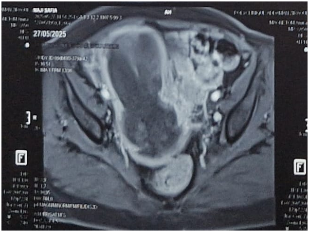
Figure 1: Pelvic MRI image showing an endocavitary uterine mass
Discussion
The management of uterine sarcoma is complex due to its rarity and high histological heterogeneity. Surgery, consisting of a total hysterectomy with bilateral adnexectomy, is the standard treatment [6]. The value of adjuvant treatments such as radiotherapy or chemotherapy remains controversial, with some studies showing limited benefit, particularly in localized forms [7]. Hormone therapy may be used in low-grade endometrial stromal sarcomas expressing hormone receptors [8]. The prognosis is generally poor: the 5-year survival rate for localized leiomyosarcoma is approximately 61%, but drops to 13% in the metastatic stage [10]. Undifferentiated sarcomas also have a poor prognosis, with an overall survival rate of around 46% [11], while low-grade endometrial stromal sarcomas reach 93% across all stages [9]. Recurrence is common, ranging from 36% to 63% depending on the series [17]. The lack of specific screening, clinical and radiological similarity to fibroids, and the aggressive nature of certain sub types explain the difficulties in management. Currentre search focuses on the identification of diagnostic and prognostic bio markers, as well as targeted therapies and immunotherapy [12].
Conclusion
Uterine sarcoma is a rare but serious condition that requires specialized, multi disciplinary care. Treatment is mainly based on surgery, but results remain limited in advanced cases. The prospects for improvement lie in earlier diagnosis, better prognostic stratification, and the integration of new therapeutic approaches, particularly targeted and immunological ones, into treatment protocols [13].
References
- Carrie Madormo. Types of Uterine Cancer. Consulté en août. Health.com. 2025. [PubMed.]
- Toro JR., Travis LB., Wu HJ., Zhu K., Fletcher CD., Devesa SS. Epidemiology of uterine sarcoma in the United States: 1973–1998. J Natl Cancer Inst. 2006;98(5):393-398. [Ref.]
- Yale Medicine. Uterine Sarcoma Overview. Consulté en août. 2025. [Ref.]
- Brooks SE., Zhan M., Cote T., Baquet CR. Surveillance, epidemiology, and end resultsanalysis of 2677 cases of uterinesarcoma 1989–1999. Gynecol Oncol. 2004;93(1):204-208. [PubMed.]
- D’Angelo E., Prat J. Uterine sarcomas: a review. Gynecol Oncol. 2010;116(1):131-139. [PubMed.]
- Morice P., Leary A., Creutzberg C., Abu-Rustum N., Darai E. Surgery in uterine sarcomas:principles and outcomes. Eur J Obstet Gynecol Reprod Biol. 2012;165(2):255-263. [Ref.]
- Hensley ML., Miller A., O’Malley DM., et al. Adjuvant therapy for high-grade uterinesarcoma: aretrospectivestudy. Gynecol Oncol. 2013;128(3):321-325. [Ref.]
- Mackay HJ., Ray-Coquard I., et al. Hormonal therapy in low-grade endometrial stromal sarcoma. Gynecol Oncol. 2012;125(1):142-146. [PubMed.]
- Endometrial stromal sarcoma survival. Consulté en août. American Cancer Society. 2025. [PubMed.]
- American Cancer Society. Survival rates for uterinesarcoma. Consulté en août. America Cancer Society. 2025. [Ref.]
- Undifferentiated uterine sarcoma survival. Consulté en août. American Cancer Society. 2025. [Ref.]
- Nathenson MJ., Ravi V., Fleming N., Wang WL., Conley AP. Uterine sarcomas: molecular features., management., and future directions. Curr Oncol Rep. 2016;18(6):41. [PubMed.]
- Scambia G., Fagotti A., Fanfani F., et al. New therapeutic strategies in uterine sarcomas. Int J Gynecol Cancer. 2020;30(9):1261-1268. [Ref.]
- Hensley ML., Chiu J., Maki R. Uterine leiomyosarcoma: management and treatment. Curr Opin Oncol. 2013;25(4):384-389. [Ref.]
- Amant F., Floquet A., Friedlander M., et al. Endometrial stromal sarcoma:clinical review and management recommendations. GynecolOncol. 2014;132(1):1-10. [Ref.]
- Cancer.gov. Uterine Carcinosarcoma Treatment (PDQ®). Consulté en août 2025. National Cancer Institute. 2025. [Ref.]
- Pautier P., Floquet A., Gladieff L., et al. Uterinesarcoma: patterns of recurrence and survival. Gynecol Oncol. 2023;168(2):290-298. [Ref.]