>Corresponding Author : Nouri Meriem
>Article Type : Case Report
>Volume : 5 | Issue : 10
>Received Date : 06 August, 2025
>Accepted Date : 12 August, 2025
>Published Date : 25 August, 2025
>DOI : https://doi.org/10.54289/JCRMH2500147
>Citation : Nouri M, Etaouas C, El Bouadi O, Jalal M, Lamrissi A, et al. (2025) Abdominal Pregnancy After Tubal Rupture: A Case Report. J Case Rep Med Hist 5(10): doi https://doi.org/10.54289/JCRMH2500147
>Copyright : © 2025 Nouri M, et al. This is an open-access article distributed under the terms of the Creative Commons Attribution License, which permits unrestricted use, distribution, and reproduction in any medium, provided the original author and source are credited.
Case Report | Open Access | Full Text
1Department of Maternity, Abderrahim El Harouchi Mother and Child Hospital, Ibn
2Rochd University Hospital Center, Faculty of Medicine and Pharmacy, Hassan II, University of Casablanca, Morocco
*Corresponding author: Nouri Meriem, Department of Maternity, Abderrahim El Harouchi Mother and Child Hospital, Ibn
Abstract
Abdominal pregnancy is a rare type of ectopic pregnancy that only accounts for less than 1% of cases. It can be primary or, more frequently, secondary to a tubal rupture. We report the case of a 22-year-old woman, first action, null, without any particular history. The patient, whose medical monitoring of pregnancy was irregular, had been consulted for acute abdominal pain with a 5-month delay in management. Laparotomy confirmed diagnosis of abdominal pregnancy. In our case, the abdominal pregnancy is secondary and due to the implantation of the egg in the peritoneal cavity following a tubal rupture. In developing countries, two factors explain the high incidence of abdominal pregnancy: high incidence of genital infections and poor pregnancy follow-up. case of abdominal pregnancy: high genital infection incidence and the poor pregnancy follow-up.
Keywords: Abdominal pregnancy, Ultrasonography, Plain abdominal radiography, Surgery
Abbreviations: EP: Ectopic Pregnancy, BP: Blood Pressure, BHCG: Beta-Human Chorionic Gonadotropin, SA: Semaines d'Aménorrhée, IVF: In Vitro Fertilization, CT: Computed Tomography
Introduction
Abdominal pregnancy is a rare entity of ectopic pregnancy with implantation of the egg in the large abdominal cavity [1]. It is a rare eventuality and the prerogative of countries with a low socio-economic level and little medical care [2]. It may be primitive, when fertilization has taken place during intra-abdominal migration of the ovum, or secondary to uterine rupture after an old corporal caesarean section, or on a malformed uterus, or to tubal pregnancy by rupture of the tube, or after migration of the fertilized egg into the abdominal cavity. This explains the need for early detection [3].
Case Report
22-year-old patient, nulliparous, with no particular pathological history, admitted to the emergency department for metrorrhagia associated with pelvic pain after 5 months of amenorrhea. Examination revealed a tachycardic patient, BP 90/50 mmhg, generalized mucocutaneous pallor, sweating, slightly distended abdomen with generalized defensiveness, bleeding from the endocervix. Pelvic ultrasound showed an empty uterus of normal size, a retrouterine gestational sac measuring 88x43mm containing a live embryo biometrically estimated to be pregnant at 15 weeks of amenorrhea, associated with a copious effusion (Figure 1), and a BHCG level of 79859. She underwent emergency laparotomy, exploration showed a hemoperitoneum estimated at 2 liters, An ovoid fetal sac measuring 10 cm, implanted directly between the uterus and the rectum, adherent to the omentum at the level of the cul de sac of the Douglas (Figure 2, Figure 3).
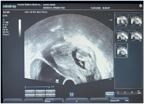
Figure 1: Transvaginal ultrasound showing a retrouterine gestational sac with a live embryo, consistent with secondary abdominal pregnancy at 15 weeks of amenorrhea.

Figure 2: The fetus and placental

Figure 3: Surgical exploration
Discussion
Atypical pregnancy is one of the most important haemorrhagic emergencies in obstetrics and gynaecology. It is a frequent cause of morbidity and sometimes mortality in women of childbearing age. Its incidence has risen steadily in recent years in both developing and developed countries [4, 5]. Abdominal pregnancy, however, is a rare variety of ectopic pregnancy (1% of all ectopic pregnancies) [6]. It is exceptional in wealthy countries (1/7,000 to 1/15,000 live births) and more frequent in Third World countries, whose etiology has not been clearly identified [5]. In Africa, the incidence of abdominal pregnancy is less rare, ranging from 0.009% in Morocco to 0.152% in Nigeria [6]. This is due to the absence of good clinical, ultrasound and biological monitoring of pregnancy [7].
Abdominal pregnancy corresponds to the implantation and development of the fertilized egg in the peritoneal cavity. It is responsible for serious maternal-fetal morbidity and mortality.
A distinction is made between 2 types of abdominal pregnancy, based on pathophysiological mechanisms [3, 8]:
o Secondary abdominal pregnancies, the most frequent, are due to tubo-abdominal abortion, rupture of a tubal pregnancy, or migration of an intrauterine pregnancy through a hysterectomy breach, uterine perforation or rudimentary horn.
o Primary pregnancies are due to implantation of the egg in the peritoneal cavity as a result of delayed ovarian uptake.
In Our case, the associated tubal rupture can be classified as a secondary abdominal pregnancy.
However, there is a classification according to term. A distinction is made between early and advanced abdominal pregnancies diagnosed after 20 days' gestation [9].
There are several clinical criteria to guide the diagnosis of an abdominal pregnancy [10]:
• digestive disorders: nausea, vomiting, sub-occlusion;
• abdominal and pelvic pain concomitant with fetal movements, with or without metrorrhagia;
• anemia with altered general condition;
• a very superficial fetus, often in an atypical high transverse position;
Sometimes, a progressive complication such as internal or externalized hemorrhage, or a toxi-infectious syndrome.
Biological tests may show anemia and increased alpha-feto-protein levels [11].
Ultrasound plays an important role in confirming the diagnosis, showing a fetus in a gestational sac outside the uterus. It also shows fetal vitality and the site of placental insertion [12].
With regard to risk factors, those for EP are generally found: infertility, intrauterine device, pregnancy after in vitro fertilization [IVF]; history of traumatic endouterine manoeuvres: voluntary termination of pregnancy by aspiration [14], uterine scar from an abdominal pregnancy with recurrence of an advanced abdominal pregnancy from the same site Anecdotally, abdominal pregnancy after hysterectomy has also been described: 29 cases reported in the literature in 1984 [13]. None of these risk factors were found in our patient. However, she is now at risk of another abdominal pregnancy.
For therapeutic management, most authors recommend laparotomy once the diagnosis has been made, irrespective of fetal status, given the unpredictable and serious nature of maternal complications arising at any term. A conservative approach is debatable from 24 SA onwards [14]. In 1990, Martin proposed a list of 7 criteria required to propose conservative treatment for abdominal a pregnancy diagnosed after 20 SA: Absence of malformation; Absence of signs of maternal or fetal decompensation; Monitoring of fetal well-being; Placental insertion in the lower abdomen; Distance from the liver and spleen; Presence of amniotic fluid around the fetus [15] Our patient was not immediately eligible for the conservative approach due to the length of the pregnancy.
In practice, if the diagnosis is made before 20 SA, termination of pregnancy will be discussed, and derogation from this attitude should remain exceptional. If the decision is taken to maintain the pregnancy, hospitalization in an appropriate facility until birth is required. The term of programmed laparotomy varies in the literature: Ombelet in a series of 38 cases in 1988 recommended 34 SA [16].
Conclusion
Ectopic pregnancy remains a frequent pathology in our working context. The introduction of endovaginal ultrasound as a means of diagnosis would improve early detection and laparoscopic management, which could have a positive impact on patient fertility.
References
- Poizat R, Lewin F. Extra-uterine pregnancy after the 5th month. Encycl Méd Chir Obstétrique. 1982;D10:50–69. [Ref.]
- Alto WA. Abdominal pregnancy. Am Fam Physician. 1990;41(1):209–14. [PubMed.]
- Drave NA, Phelan J. Retrospective study of maternal mortality in the gynecology and obstetrics department of the National Hospital of Point G from 1991 to 1994: about 103 cases [Medical thesis]. Bamako: University of Bamako; 1996. [Ref.]
- Atrash HK, Friede REW, Hogue CJR. Abdominal pregnancy in the United States: frequency and maternal mortality. Obstet Gynecol. 1987;69(3 Pt 1):333–7. [Ref.]
- Diouf A, Diouf F, Cisse CT, Diaho D, Gaye A, Diadhiou F. Term abdominal pregnancy with live fetus: about two cases. J Gynecol Obstet Biol Reprod (Paris). 1996;25(2):212–5. [PubMed.]
- Ferland RJ, Chadwick DA, O’Brien JA, Granai CO III. An ectopic pregnancy in the upper retroperitoneum following in vitro fertilization and embryo transfer. Obstet Gynecol. 1991;78(3 Pt 2):544–6. [Ref.]
- Martin JN Jr, Sessums JK, Martin RW, Pryor JA, Morrison JC. Abdominal pregnancy: current concepts of management. Obstet Gynecol. 1988;71(4):549–57. [PubMed.]
- Shumway JB, Greenspoon JS, Khouzami AN, Platt LD, Blakemore KJ. Amniotic fluid alpha-fetoprotein (AFAFP) and maternal serum alpha-fetoprotein (MSAFP) in abdominal pregnancies: correlation with extent and site of placental implantation and clinical implications. J Matern Fetal Med. 1996;5(3):120–3. [PubMed.]
- Neiger R, Weldon K, Means N. Intramural pregnancy in a cesarean section scar: a case report. J Reprod Med. 1998;43(11):999–1001. [PubMed.]
- Costa SD, Presley J, Bastert G. Advanced abdominal pregnancy. Obstet Gynecol Surv. 1991;46(8):515–25. [PubMed.]
- Martin JN Jr, McCaul JF IV. Emergent management of abdominal pregnancy. Clin Obstet Gynecol. 1990;33(3):438–47. [PubMed.]
- Ombelet W, Vandermerwe JV, Van Assche FA. Advanced extra-uterine pregnancy: description of 38 cases with literature survey. Obstet Gynecol Surv. 1988;43(7):386–97. [PubMed.]
- Ruptured abdominal pregnancy: report of a case. N Z Med J. 1990;103(891):158–9. [Ref.]
- Abdominal pregnancy: imaging and management. Eur J Radiol. 1995;20(1):23–6. [Ref.]
- Scholar Google Citations – user: LnIxSs4AAAAJ. [Ref.]
- Bouyer J, Coste J, Fernandez H, Pouly JL, Job-Spira N. Sites of ectopic pregnancy: a 10-year population-based study of 1800 cases. Eur J Obstet Gynecol Reprod Biol. 2002;105(2):180–6. [PubMed.]