>Corresponding Author : Benhaddouga Khadija
>Article Type : Case Report
>Volume : 5 | Issue : 9
>Received Date : 21 July, 2025
>Accepted Date : 30 July, 2025
>Published Date : 12 August, 2025
>DOI : https://doi.org/10.54289/JCRMH2500144
>Citation : Benhaddouga K, Moustatir M, Ckeikh IN, Chyate FZ, Boufettal H, et al. (2025) Villo-Glandular Carcinoma of the Cervix: 1 Case Report and Review of the Literature. J Case Rep Med Hist 5(9): doi https://doi.org/10.54289/JCRMH2500144
>Copyright : © 2025 Khadija B, et al. This is an open-access article distributed under the terms of the Creative Commons Attribution License, which permits unrestricted use, distribution, and reproduction in any medium, provided the original author and source are credited.
Case Report | Open Access | Full Text
1Resident Physician, Department of Gynecology and Obstetrics, at Ibno Rochd University Hospital, Casablanca, Morocco
2Professor in the Department of Gynecology and Obstetrics at the Ibno Rochd University Hospital in Casablanca, Morocco
*Corresponding author: Benhaddouga Khadija, Resident Physician, Department of Gynecology and Obstetrics, Ibno Rochd University Hospital, Casablanca, Morocco
Abstract
Villo-glandular carcinoma of the cervix is a rare and distinct form of cervical adenocarcinoma. It is characterized by villo-papillary architecture and well-defined glandular differentiation. This entity, often seen in young women, is generally associated with a better prognosis than other types of cervical adenocarcinoma. Nevertheless, due to its rarity, its diagnosis poses a challenge, requiring close correlation between clinical, histopathological and immunohistochemical data. This review summarizes current knowledge on the epidemiology, pathogenesis, morphological features, differential diagnosis and therapeutic options of this unusual tumor.
Keywords: Villo-Glandular Carcinoma, Cervix, Adenocarcinoma, Histopathology, Prognosis
Introduction
Villo-glandular carcinoma of the cervix is a rare variant of cervical adenocarcinoma, first described by Young and Scully in 1989 [1]. This entity accounts for less than 5% of cervical adenocarcinomas and typically occurs in young women, often nulliparous or of childbearing age [2]. It is distinguished from conventional forms by its papillary architecture, well-preserved glandular differentiation and low mitotic activity, all of which contribute to a better clinical prognosis [3]. Despite its relatively benign histological presentation, failure to recognize this tumor can lead to excessive or inappropriate management. It is therefore crucial to recognize its histological and biological particularities in order to optimize treatment and avoid over-treatment.
Case Report
This is a 46-year-old patient, mother of 3 children by vaginal delivery, with no particular pathological history, presenting with post-coital metrorrhagia with dyspareunia dating back 5 months prior to her consultation. Clinical examination: normal cervix, no bleeding (figure 1).
Cervico-uterine smear: ASCUS
A colposcopy was performed, revealing a lesion at 15 h, biopsied in favour of an epithelial proliferation of villeous and glandular architecture, initially suggestive of villo-glandular carcinoma.
Pelvic MRI: no abnormalities (figure 2).
An enlarged hysterectomy without adnexal preservation was performed. Anatomopathological study revealed an infiltrating villo-glandular adenocarcinoma of the uterine cervix, 3mm thick and 8mm wide.

Figure 1: Image of the cervix
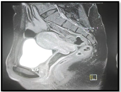
Figure 2: IRM pelvienne sans anomalie
Discussion
The origin of villo-glandular carcinoma of the cervix remains open to debate, although an association with high-risk HPV viruses has been demonstrated in some cases, notably HPV 18 [4]. Clinically, patients often present with metrorrhagia or unusual vaginal discharge, but some cases are diagnosed incidentally on cervical smear or directed biopsy [5].
Histologically, this tumor is characterized by a complex papillary architecture, sometimes arboriform, covered by a cylindrical mucosecretory epithelium, often pseudostratified [6]. Cytology is generally moderately atypical, with little mitotic activity. These features can make it difficult to distinguish from benign lesions such as villo-papillary adenoma or glandular papilloma, sometimes requiring immunohistochemistry to assess expression of p16, Ki-67 or the progesterone receptor [7].
Differential diagnosis should include classic endocervical adenocarcinomas, endometrioid carcinoma and metastatic tumours with a glandular component. A useful feature is the preservation of the basement membrane and the absence of deep stromal invasion, especially in early forms [8].
Treatment of villo-glandular carcinoma depends on tumour stage and patient age. Given its predominance in younger women, a Conservative approach may be considered in early forms, particularly in the absence of lymphovascular invasion [9]. Wide conization with healthy margins may suffice in vertlocalized cases, while hysterectomy remains indicated in more advanced stages or in cases of recurrence [10].
The prognosis of this tumor is generally favorable. Several studies have shown a low incidence of lymph node metastases and a 5-year survival rate in excess of 90% in early stages [11]. However, rare cases of aggressive forms with deep invasion or extrapelvic extension have been reported, underlining the need for prolonged and rigorous follow-up [12].
Conclusion
Villo-glandular carcinoma of the cervix is a rare but distinct tumor entity, with specific morphological and clinical features. Its recognition is essential to avoid overtreatment, especially in young patients. Diagnosis is based on careful histological evaluation, supported where necessary by immunohistochemistry. Although its prognosis is generally good, close surveillance is necessary due to its potentially invasive course in certain cases.
References
- Villo-glandular carcinoma of the cervix is a rare but distinct tumor entity, with specific morphological and clinical features. Its recognition is essential to avoid overtreatment, especially in young patients. Diagnosis is based on careful histological evaluation, supported where necessary by immunohistochemistry. Although its prognosis is generally good, close surveillance is necessary due to its potentially invasive course in certain cases. [PubMed.]
- Jones MW., et al. Papillary adenocarcinoma of the uterine cervix: a clinicopathologic study of 36 cases. Int J Gynecol Pathol. 1993;12(1):1–7. [Ref.]
- Silver SA., Devouassoux-Shisheboran M., Mezzetti TP., et al. Papillary adenocarcinomas of the uterine cervix. Mod Pathol. 2000;13(8):951–956. [Ref.]
- Park JY., et al. HPV typing in villoglandular papillary adenocarcinoma. Gynecol Oncol. 2002;86(3):239–244. [Ref.]
- Tringali S., et al. Villoglandular adenocarcinoma of the uterine cervix: diagnostic and management issues. J Low Genit Tract Dis. 2013;17(2):221–225. [Ref.]
- Ishii K., et al. Histological and cytological characteristics of villoglandular adenocarcinoma. Acta Cytol. 2004;48(5):587–592. [Ref.]
- Talia KL., et al. Immunoprofile of villoglandular adenocarcinoma. Pathology. 2016;48(2):171–177. [Ref.]
- McCluggage WG. New developments in endocervical glandular lesions. Histopathology. 2013;62(1):138–160. [PubMed.]
- Bisseling KC., et al. Conservative treatment of early stage villoglandular papillary adenocarcinoma. Obstet Gynecol. 2007;109(2 Pt 2):463–466. [Ref.]
- Schnatz PF., et al. Villoglandular adenocarcinoma of the cervix: a systematic review. Obstet Gynecol Surv. 2010;65(6):389–395. [Ref.]
- Utsugi K., et al. Clinical behavior of villoglandular adenocarcinoma. J Obstet Gynaecol Res. 2012;38(9):1206–1211. [Ref.]
- Wang X., et al. Aggressive variant of villoglandular adenocarcinoma. Int J Gynecol Pathol. 2015;34(6):560–565. [Ref.]