>Corresponding Author : Benhaddouga Khadija
>Article Type : Case Report
>Volume : 5 | Issue : 8
>Received Date : 06 July, 2025
>Accepted Date : 16 July, 2025
>Published Date : 20 July, 2025
>DOI : https://doi.org/10.54289/JCRMH2500137
>Citation : Benhaddouga K, Mazhour M, Nejat CI, Medyani H, Bensouda M, et al. (2025) Spina Bifida: 1 Case Report and Review of the Literature. J Case Rep Med Hist 5(8): doi https://doi.org/10.54289/JCRMH2500137
>Copyright : © 2025 Benhaddouga K, et al. This is an open-access article distributed under the terms of the Creative Commons Attribution License, which permits unrestricted use, distribution, and reproduction in any medium, provided the original author and source are credited.
Case Report | Open Access | Full Text
1Resident Physician, Department of Gynecology and Obstetrics, at Ibno Rochd University Hospital, Casablanca, Morocco
2Professor in the Department of Gynecology and Obstetrics at the Ibno Rochd University Hospital in Casablanca, Morocco
*Corresponding author: Benhaddouga Khadija, Resident Physician, Department of Gynecology and Obstetrics, at Ibno Rochd University Hospital, Casablanca, Morocco
Abstract
Spina bifida is a congenital malformation of the neural tube, affecting the spinal column and sometimes the spinal cord. This pathology can lead to varying degrees of motor, sensory and cognitive disability, depending on the severity of the lesion. Prenatal diagnosis, folic acid supplementation, early surgical intervention and multidisciplinary management have improved patients' prognosis and quality of life.
Keywords: Spina bifida, Neural tube defect, Folic acid, Disability, Prenatal Surgery, Myelomeningocele
Abbreviations: SA: Semaines d’Aménorrhée, CNS: Central Nervous System, AFP: Alpha-Fetoprotein, CS: Caesarean Section, MRI: Magnetic Resonance Imaging, NTD: Neural Tube Defect, Chiari II: Chiari Type II Malformation
Introduction
Spina bifida is a neural tube defect that occurs during the first weeks of gestation. It is one of the most frequent congenital anomalies of the central nervous system. Its prevalence varies according to region and prevention policies, notably folic acid supplementation. Three main forms are recognized: spina bifida occulta, meningocele and myelomeningocele, the latter being the most severe and disabling. Early recognition and appropriate management are essential to limit functional complications.
Case Report:
The patient was 39 years old, mother of a vaginal birth, with no history of consanguinity, and no particular pathological history of a 39SA pregnancy
On admission, the patient was conscious, hemodynamically and respiratory stable. Urine dipstick negative.
Obstetrical ultrasound: progressive monofetal pregnancy, positive cardiac activity with ultrasound appearance of major hydrocephalus + Spina bifida
The patient gave birth to a male neonate, apgar 8/10, birth weight 3900g by caesarean section. (figure 1)
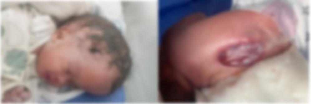
Figure 1: Hydrocephalus + spina bifida
Discussion
Spina bifida results from a defect in the closure of the posterior part of the neural tube, usually between days 22 and 28 after conception. Several factors are involved, including genetic, environmental and nutritional factors, in particular folic acid deficiency during the periconceptional period (1). Prevention is therefore largely based on systematic folic acid supplementation in women of childbearing age (2). Despite this, cases continue to occur, justifying the importance of prenatal screening by ultrasound and determination of alpha-fetoprotein in amniotic fluid.
Clinical forms vary: the occult form is often asymptomatic, while myelomeningocele manifests as a hernia containing meninges and spinal cord through a vertebral opening, causing flaccid paralysis, urinary and fecal incontinence, as well as orthopedic disorders (3). Diagnosis may be made in utero or at birth. Associated malformations, notably hydrocephalus and Chiari type II malformation, are common and worsen the prognosis (4).
The treatment of spina bifida is multidisciplinary. Neonatal surgery or, in certain specialized centers, fetal surgery can close the lesion and reduce the risk of infection. Prenatal surgery, though complex, has been shown to improve motor function and reduce the incidence of hydrocephalus (5). However, it also entails significant obstetrical risks. Postnatal management includes neurosurgery, rehabilitation, urological management, orthopedics and psychosocial follow-up.
Patients' quality of life depends on the severity of the malformation, the level of injury, early care and family and institutional support. Thanks to medical advances, many children with spina bifida can reach adulthood with a good level of autonomy, although sequelae remain frequent (6). School support and social integration are also challenges to be met in order to promote their inclusion.
Conclusion
Spina bifida remains a serious condition with multiple repercussions, but advances in diagnosis and treatment have improved its outcome. Primary prevention with folic acid, prenatal screening, early surgical approaches and coordinated management are the pillars of the management of this malformation. Greater awareness among healthcare professionals and the general public is still needed to reduce its incidence and improve patients' quality of life.
Reference
- Czeizel AE, Dudás I. Prevention of the first occurrence of neural-tube defects by periconceptional vitamin supplementation. N Engl J Med. 1992;327(26):1832–5. [PubMed.]
- Botto LD, Lisi A, Bower C, et al. Trends of selected malformations in relation to folic acid recommendations and fortification: an international assessment. Birth Defects Res A Clin Mol Teratol. 2006;76(10):693–705. [PubMed.]
- Copp AJ, Greene ND. Genetics and development of neural tube defects. J Pathol. 2010;220(2):217–30. [PubMed.]
- Dias MS, Partington MD. Embryology of myelomeningocele and anencephaly. Neurosurg Focus. 2004;16(2): E1. [PubMed.]
- Adzick NS, Thom EA, Spong CY, et al. A randomized trial of prenatal versus postnatal repair of Myelomeningocele. N Engl J Med. 2011;364(11):993–1004. [PubMed] [PubMed.]
- Bowman RM, McLone DG, Grant JA, Tomita T, Ito JA. Spina bifida outcome: a 25-year prospective. Pediatr Neurosurg. 2001;34(3):114–20. [PubMed.]